Veterinary ultrasound is one of the most important diagnostic tools in modern animal healthcare, second only to radiography in terms of widespread use. It is widely used in both large and small animals for assessing internal organs, detecting pregnancy, diagnosing musculoskeletal injuries, and evaluating cardiovascular function. The principle behind this technology lies in the use of high-frequency sound waves to produce real-time images of internal body structures. This article explores the working principles of veterinary ultrasound, its various imaging modes, applications in different species, and recent advancements that are transforming veterinary diagnostics.
Basic Principle of Veterinary Ultrasound
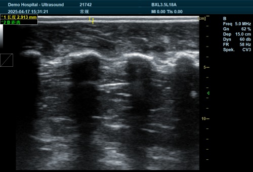
At the core of veterinary ultrasonography is the transmission and reception of sound waves. A device called a transducer generates ultrasonic pulses, usually in the frequency range of 1.5–18 megahertz (MHz). These sound waves travel into the animal’s body and encounter various tissues and organs, each with a different density. When the sound waves hit a boundary between tissues of different densities, part of the wave is reflected back to the transducer as an echo, while the rest continues deeper into the body.
The transducer then switches into a receiving mode to collect the returning echoes. These echoes are converted into electrical signals and processed by the ultrasound machine’s computer to generate an image. The image represents a “slice” of the animal’s internal structures, allowing the veterinarian to observe organ shape, size, and composition in real-time.
Key Imaging Modes
Several types of imaging formats are used in veterinary ultrasound:
-
B-mode (Brightness mode): This is the most commonly used imaging mode. It creates a two-dimensional grayscale image representing the anatomical structure. Each echo is displayed as a dot, and its brightness reflects the strength of the echo.
-
M-mode (Motion mode): Primarily used in cardiac imaging (echocardiography), this mode records the motion of organs, such as heart valves, over time along a single line of ultrasound.
-
Doppler mode: This mode evaluates blood flow by using the Doppler effect, which measures changes in frequency of sound waves reflected from moving blood cells. It helps detect abnormalities like valve regurgitation, vessel narrowing, and irregular blood flow patterns.
-
Color Doppler: This version of Doppler mode overlays color-coded blood flow direction and velocity on the B-mode image, offering a more comprehensive view of circulatory function.
Image Formation and Real-Time Scanning
Modern ultrasound machines produce real-time images by rapidly sweeping the sound beam through the body many times per second. This creates a moving image that changes as the transducer is shifted across the skin. A transmission gel is applied between the transducer and the skin to ensure optimal sound wave conduction.
The system records not only the strength of each echo but also the time it takes to return and the direction of the sound beam. These details are essential for accurately reconstructing the internal layout of the scanned tissues.

Limitations and Challenges
Despite its advantages, ultrasound has some limitations:
-
Limited penetration depth: High-frequency probes provide high-resolution images but cannot penetrate deeply. Lower-frequency probes can reach deeper structures but with less detail.
-
Gas and bone interference: Ultrasound waves do not travel well through air or dense bone. Gas in the intestines and bones can block or distort the image, limiting visibility of deeper structures.
-
Noise and artifact formation: At greater depths (e.g., >30 cm), echoes become weaker and are harder to distinguish from background electronic noise.
Common Applications in Veterinary Practice
Reproductive Health
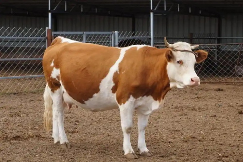
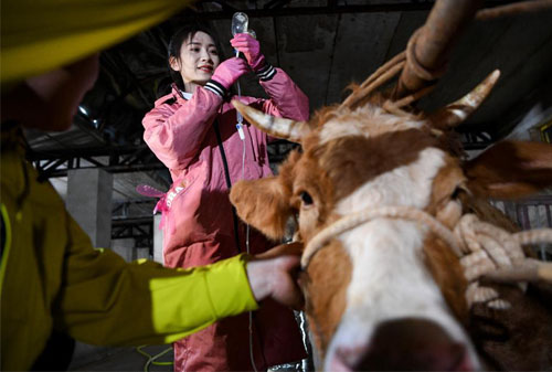
Ultrasound is widely used in reproductive management, particularly in large animals. Transrectal ultrasonography allows veterinarians to monitor ovarian follicles, detect pregnancy, and diagnose reproductive disorders in cows and mares.
Musculoskeletal System
In horses and dogs, ultrasound is often used to evaluate tendon and ligament injuries. This non-invasive method provides detailed images of soft tissue structures that cannot be easily seen on X-rays.
Abdominal Imaging
Veterinarians use ultrasound to examine abdominal organs such as the liver, spleen, kidneys, bladder, and intestines. It helps detect tumors, cysts, and obstructions, as well as evaluate organ size and texture.
Cardiovascular Assessment (Echocardiography)
Ultrasound of the heart (echocardiography) provides valuable information about cardiac anatomy and function. Using a combination of B-mode, M-mode, and Doppler ultrasound, veterinarians can measure chamber size, wall motion, valve function, and blood flow patterns.
Advances in Ultrasound Technology
Recent developments have significantly enhanced the utility of veterinary ultrasound:
-
3D and 4D imaging: These technologies allow for volumetric imaging and real-time motion of structures in three dimensions, improving diagnostic accuracy.
-
Elastography: This technique evaluates tissue stiffness, which can help differentiate between benign and malignant lesions.
-
Contrast-enhanced ultrasound (CEUS): CEUS uses microbubble contrast agents to enhance the visibility of blood vessels and improve the detection of certain types of tumors and organ perfusion.
Ultrasound-Guided Procedures
Ultrasound is also used to guide needle biopsies and fluid aspirations, increasing accuracy and reducing the risk of complications. It enables veterinarians to collect tissue samples from deep or delicate areas without the need for exploratory surgery. This technique is particularly helpful for diagnosing conditions affecting the liver, kidneys, lymph nodes, and abdominal masses.
Importance of Experience and Interpretation
Accurate interpretation of ultrasound images requires training and experience. The echogenicity (brightness) of tissues varies depending on the organ, its composition, and the type of transducer used. Comparison with normal anatomical patterns is essential for identifying abnormalities. Often, subtle changes in tissue structure or blood flow provide critical clues that only experienced practitioners can recognize.
Conclusion
Veterinary ultrasound is an indispensable tool in modern animal health care, offering a non-invasive, safe, and efficient way to examine internal structures. Its ability to provide real-time images, assess soft tissue, and guide diagnostic procedures makes it a preferred choice for many clinical applications. While it has limitations in certain conditions—such as gas interference and limited depth penetration—ongoing technological advancements are continually expanding its diagnostic capabilities. For both livestock and companion animals, veterinary ultrasound plays a vital role in early detection, accurate diagnosis, and effective treatment planning.
Reference
-
American College of Veterinary Radiology. (n.d.). Ultrasound in Veterinary Medicine. Retrieved from https://acvr.org
-
Veterinary Imaging Center of San Diego. (n.d.). Understanding Veterinary Ultrasound. Retrieved from https://www.vicsd.com
-
PennVet. (n.d.). Veterinary Diagnostic Imaging Services. Retrieved from https://www.vet.upenn.edu