Veterinary ultrasound is one of the most essential and widely used diagnostic imaging tools in animal healthcare today. Utilizing high-frequency sound waves—usually between 1.5 to 18 MHz—this non-invasive technology allows veterinarians to visualize internal organs and tissues in real-time without the need for surgery. Its applications span across species and specialties, helping to diagnose, monitor, and guide treatments for various conditions. In this article, we will explore what veterinary ultrasound machines can detect, how they work, and why they are indispensable in modern animal care.

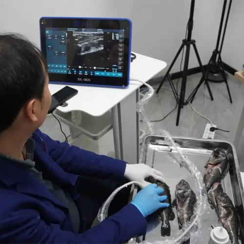
Fish Ultrasound Images
How Veterinary Ultrasound Works
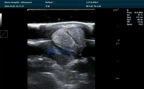
Ultrasound imaging, also known as ultrasonography, operates on the principle of sound wave reflection. A transducer sends sound pulses into the body, and when these waves encounter tissues of different densities, they reflect back at varying intensities. The ultrasound machine then processes these echoes to create detailed images of internal structures.
The most common imaging mode used is B-mode (brightness mode), which produces a grayscale image representing the anatomy. In veterinary practice, real-time scanning allows veterinarians to observe moving structures—such as a beating heart or fetal movement—and adjust the transducer to obtain the best angle and clarity.
Structures and Conditions Detected by Veterinary Ultrasound
1. Abdominal Organs
Ultrasound is particularly effective for assessing soft tissues in the abdomen. It can detect changes in size, shape, texture, and echogenicity (brightness) of organs such as:
-
Liver: Inflammation, tumors, cysts, and bile duct obstructions
-
Kidneys: Stones, cysts, chronic disease, and tumors
-
Spleen: Enlargement, masses, or rupture
-
Bladder: Stones, tumors, wall thickening, and residual urine
-
Intestines: Blockages, foreign bodies, thickening, or inflammation
Although gas in the intestines can limit visibility, strategic scanning angles and fasting the animal beforehand can improve results.
2. Reproductive System
In both large and small animals, ultrasound plays a vital role in reproductive management:
-
Pregnancy diagnosis: Detects fetal heartbeat and viability
-
Ovarian and uterine evaluation: Assesses follicle development, cysts, infections
-
Testicular examination: Detects tumors, abscesses, or inflammation
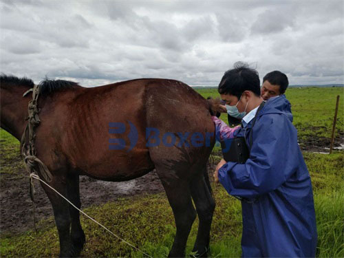
In large animals such as cattle or horses, transrectal ultrasound is frequently used to monitor breeding status or guide artificial insemination and embryo transfer procedures.
3. Musculoskeletal System
Ultrasound can evaluate muscles, tendons, ligaments, and joints, especially in performance animals such as horses and working dogs.
-
Tendon injuries: Detects tears, strains, and healing progression
-
Joint assessment: Identifies effusion, capsule thickening, cartilage damage
-
Bone surfaces: Assesses periosteal reactions and joint margins
In equine medicine, ultrasound is a first-line imaging modality for lameness evaluations due to its portability and safety.
4. Cardiac Imaging (Echocardiography)
Echocardiography is a specialized ultrasound application to assess the heart’s structure and function.
-
M-mode: Captures motion over time, useful for evaluating valve movement and chamber dimensions
-
B-mode: Provides cross-sectional anatomical views
-
Doppler ultrasound: Evaluates blood flow direction and speed, helping diagnose valve regurgitation, stenosis, or congenital defects
This technique is crucial for diagnosing heart diseases, monitoring progression, and guiding treatment in both large and small animals.
Advanced Applications of Veterinary Ultrasound
1. Ultrasound-Guided Procedures
Ultrasound allows veterinarians to perform minimally invasive procedures with high precision:
-
Fine-needle aspiration (FNA): Collects cells from masses or organs
-
Biopsies: Extracts tissue samples from liver, kidney, or lymph nodes
-
Fluid drainage: Removes fluid from cysts, abscesses, or thoracic cavities
These methods reduce the need for exploratory surgery and provide quicker diagnoses with fewer complications.
2. Emergency and Critical Care
In emergency settings, ultrasound can rapidly identify life-threatening conditions such as:
-
Free fluid in the abdomen or chest (effusion)
-
Organ rupture or bleeding
-
Gastrointestinal dilation (e.g., gastric torsion)
FAST (Focused Assessment with Sonography for Trauma) exams are commonly performed in critical care scenarios to evaluate internal bleeding.
3. Pancreas and Endocrine Organs
Modern ultrasound machines can now detect subtle changes in organs like the pancreas and adrenal glands, which were historically difficult to visualize.
-
Pancreatitis: Identified by swelling, echogenicity changes, or peripancreatic fluid
-
Adrenal abnormalities: Including tumors, hyperplasia, or nodular changes
Though these findings often need to be confirmed with lab tests or other diagnostics, ultrasound offers a strong starting point.
Limitations of Veterinary Ultrasound
Despite its versatility, ultrasound has limitations:
-
Poor penetration through gas or bone: Lungs and bones obscure deeper structures
-
Depth limitations: Deep tissues may not reflect enough sound waves for clear imaging
-
Operator dependency: Image quality and interpretation heavily depend on the examiner’s skill and experience
Different transducers (probes) are selected based on the depth and resolution needed, with low-frequency probes offering deeper penetration but lower resolution.
Doppler Ultrasound: Seeing Blood Flow
Doppler ultrasound adds a new dimension to veterinary imaging by visualizing blood flow.
-
Spectral Doppler: Measures flow velocity at a specific site, providing waveforms over time
-
Color Doppler: Maps flow direction and speed with color coding—blue for flow toward and red for flow away from the probe
This technique is invaluable for assessing vascular patency, heart valve function, and identifying abnormal flow patterns in arteries and veins.
Conclusion: A Powerful Diagnostic Ally
Veterinary ultrasound machines are capable of detecting a wide range of conditions—from common issues like bladder stones and pregnancies to complex diseases such as pancreatitis or heart valve dysfunction. Their non-invasive nature, real-time imaging, and portability make them invaluable in both routine and emergency veterinary care.
As technology continues to advance, ultrasound devices are becoming more compact, accurate, and accessible. For veterinarians, mastering ultrasonography means offering better diagnostics, faster treatment decisions, and improved animal welfare across species and settings.