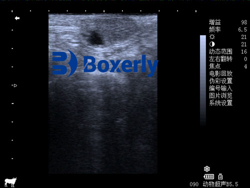
In modern cattle operations, precise reproductive management is crucial for profitability. Portable ultrasound has become a standard farm technology for this purpose. Unlike clinic machines of the past, today’s handheld scanners are small, rugged, and battery-powered, enabling accurate on-farm scanning. We will explore how portable ultrasound is used on the farm to assess cow fertility and guide breeding decisions. For dairy and beef producers, optimizing pregnancy rates directly boosts profit: each extra day a cow remains open costs feed and labor, so early detection of empty cows is economically critical. Even a modest improvement in conception rate (for example, raising the 21-day pregnancy rate by a few points on a 1,000-cow herd) can save tens of thousands of dollars annually. As a result, many farmers now integrate ultrasound into breeding programs (alongside estrus synchronization and AI) to make decisions more data-driven and timely.

Early Pregnancy Detection
Portable ultrasound excels at early pregnancy diagnosis. Skilled operators can confirm pregnancy by about 25–30 days after insemination through transrectal scanning. This is much earlier than the 35+ days needed for confident manual palpation and earlier than most hormone tests. Veterinarians report that fetal heartbeat is often visible by day 21 and that ultrasound yields near-100% accuracy by day 28. Most portable scanners use 5–7 MHz linear probes and are inserted rectally. The process is quick: after brief preparation (applying gel and positioning the probe), a trained technician can scan multiple cows in minutes. Modern devices often record images and allow data to be wirelessly saved, seamlessly adding results to herd reproductive records.
-
Rapid results: Ultrasound gives instant on-screen confirmation of pregnancy status, whereas blood or milk tests require sample analysis off-site (taking days to return results). Early detection means empty cows can be rebred sooner, saving weeks of feed and time.
-
High accuracy: As noted, ultrasound screening achieves nearly 100% accuracy by day 28 in experienced hands, reducing the chance of misdiagnosis compared to palpation.
-
Extra insight: A single ultrasound scan can reveal twins, show fetal heartbeat, and estimate fetal age. Hormone tests only indicate pregnancy presence or absence, whereas ultrasound provides detailed anatomical information immediately.
Ovarian and Uterine Evaluation
Ultrasound reveals ovarian and uterine health, both of which critically affect fertility. By scanning the ovaries, we visualize developing follicles and corpora lutea (CL). The CL is especially important: a healthy, solid CL produces progesterone that supports pregnancy. In practice, cows with a fluid-filled (“cavitary”) CL at pregnancy check are far more likely to fail conceiving. One large study found cows with a cavitary CL had 21× higher odds of remaining open than cows with a normal CL. Conversely, ultrasound-detected ovarian cysts severely impair fertility: that same study showed an ultrasound-detected cyst decreased pregnancy odds by 81-fold.
We often use ultrasound scans to guide clinical decisions. For example, if a cow shows uterine fluid or retained placenta on scan, we may delay breeding and focus on treatment first. We can also detect early embryonic loss: a follow-up scan 2–3 weeks after breeding will show whether a viable embryo is present. If the embryo is gone, the cow can be rebred immediately instead of waiting for her to return to heat. Catching these losses quickly prevents wasted estrous cycles and keeps cows on schedule.
Typical ultrasonographic assessments include:
-
Corpus luteum (CL): Evaluate size and texture. A robust, solid CL usually indicates a normal cycle; a fluid-filled (cavitary) CL often signals trouble.
-
Follicle tracking: Monitor dominant follicle growth to optimally time insemination or diagnose follicular cysts.
-
Ovarian cysts or masses: Identify any cysts or tumors on the ovaries or in the uterus. (As noted, cysts dramatically reduce conception chances.)
-
Uterine involution: Measure uterine horn diameter and wall thickness postpartum. Normal cows show rapid involution (shrinking) by a few weeks after calving, which we confirm on ultrasound. This correlates with the cow resuming cycles. Delayed involution or abnormal fluid suggests infection.
-
Twin diagnosis: Scan early to distinguish singleton vs. twin pregnancy. Detecting twins in advance informs nutrition and calving management plans.
Other useful applications:
-
Infection detection: Ultrasound can reveal uterine inflammation (fluid and debris), aiding diagnosis of metritis or endometritis so that treatment can begin early.
-
Growth monitoring: Repeated scans on young stock or yearlings verify that organs and body condition are developing normally over time.
-
Fetal sexing: By about 60–90 days of gestation, trained operators often determine fetal sex via ultrasound. This assists in planning (for example, raising mostly heifers for replacements).
-
Herd pregnancy rate: Scanning an entire bred group around 30 days post-insemination provides an accurate herd pregnancy rate and identifies cows needing rebreeding.

Bull Fertility Evaluation
Portable ultrasound also aids bull fertility exams. Although routine breeding soundness exams (BSE) often skip imaging, scanning a bull’s testes and accessory glands can uncover hidden problems. Veterinarians report using portable scanners on bulls with infertility issues: for example, any bull with low semen volume, pyospermia (pus in semen), or many abnormal sperm might be scanned for lesions. The same portable B-mode unit used for cows can image the bull’s scrotum and pelvis.
Key ultrasound checks in bulls include:
-
Testicular structure: Evaluate each testis for size, symmetry, texture, and presence of fluid. Detect lesions, tumors, or degenerative changes that might impair sperm production.
-
Epididymis and accessory glands: Scan the epididymis and prostate for inflammation or blockages affecting sperm transport.
-
Comparative findings: Ultrasound can reveal subtle pathology that routine palpation or semen analysis might miss.
As experts note, ultrasound offers a “major advantage” to identify and assess morphological changes in the bull’s reproductive tract. With more veterinarians trained in sonography, portable units are increasingly used to diagnose bull fertility issues and improve breeding soundness evaluations.
Advantages of Portable Ultrasound
Modern portable ultrasound machines offer many practical advantages in fertility management:
-
Non-invasive and Safe: Scanning is done rectally (for cows) or externally (for bulls) with no surgery or radiation. The process is gentle, causing minimal stress to animals. Veterinarians note that ultrasound is much easier on cows (and on the vet’s arms) than forceful manual palpation.
-
Immediate, Detailed Feedback: The live image reveals pregnancy and pathology instantly, allowing prompt action. We see not only pregnancy status but also fetal heartbeat, gestational age, and reproductive tract details in real time.
-
Faster Breeding Decisions: By identifying open cows early, producers can rebreed them quickly. This shortens the calving interval and increases the number of calves per cow. Many producers find they can schedule re-insemination or culling on the same day as the ultrasound scan.
-
Cost Savings: Although a quality portable unit requires investment, it often pays off quickly. For example, a Bavarian dairy cooperative reported a 14% improvement in calving intervals and a 20% drop in open cows after training technicians to do on-farm ultrasound checks. By comparison, repeatedly feeding empty cows or missing breedings can cost far more. Portable scanning also reduces vet fees (routine checks can be done by farm staff).
-
Enhanced Data: Ultrasound yields quantitative measurements (e.g. CL area, follicle diameter, uterine thickness) that can be tracked over time. This data helps fine-tune nutrition and breeding protocols. Over successive cycles, we build a database on each cow’s fertility profile.
-
Improved Welfare: Ultrasound is gentler and faster for cows than prolonged palpation or rectal exams. This improves animal welfare and reduces injury risk. It also reduces strain on farm workers and veterinarians.
-
Producer Empowerment: Many progressive farmers now buy their own scanners. This allows pregnancy checks on their schedule, integrating scanning into daily routines. It gives them confidence and control over breeding decisions without waiting for a veterinarian’s visit.
-
Versatility: Some portable units and probes can be used across species (cattle, horses, sheep), making them a multi-use farm investment. Additional modes like color Doppler also enable blood flow assessments in more advanced applications.
-
Digital Recordkeeping: Modern scanners can save images and videos. These records can be reviewed later or shared with specialists if any doubt arises. Having an archive of scans adds another layer of documentation to herd health records.
Global Adoption and Trends
Portable ultrasound has rapidly become an integral tool worldwide. In seasonal-calving systems (e.g. New Zealand, Ireland), early pregnancy checks via ultrasound are standard practice, allowing farmers to identify open cows before the next breeding season and substantially improve herd fertility. In intensive breeding programs, ultrasound data (such as ovarian function and embryo development) is even incorporated into genetic selection decisions. For instance, European and North American breeding programs use CL and follicle monitoring to refine reproductive protocols.
Training and technology advances are synergistic. Programs like “VetScan for Farmers” in Australia teach producers basic scanning so they need not wait for vets. Many veterinary services now sell or rent portable units. Hardware is also improving: new scanners offer waterproof designs, long battery life, wireless connectivity and smartphone integration. Prices continue to fall, making the technology accessible to smaller operations as well.
Limitations and Considerations
Portable ultrasound is powerful but requires proper use. The main challenge is operator skill: inaccurate interpretation can lead to errors (a small embryo might be missed or an artifact misread). Adequate training and practice are crucial. Portable probes may have slightly lower penetration than high-end consoles, so very deep imaging in obese cattle can be challenging.
Another consideration is biosecurity. Equipment must be disinfected between animals or farms to avoid disease spread. On the other hand, using a personal scanner on each farm can actually reduce infection risk compared to borrowing equipment from others, a benefit noted by producers.
There is also the initial cost: even “low-cost” units represent a significant investment. However, in herds where timely pregnancy diagnosis is critical, most managers find the return justifies the expense. Portable ultrasound does not replace good genetics, nutrition, or heat detection, but it complements them. In practice, ultrasound used alongside hormonal programs and record analysis yields the best results.
Conclusion
Bovine fertility underpins the entire cattle industry: without successful breeding, there are no calves and no product. Portable ultrasonography has revolutionized fertility assessment by providing an accurate, real-time window into each cow’s reproductive status. This technology enables much earlier pregnancy confirmation, identification of ovarian or uterine problems, and even bull fertility evaluation. Armed with ultrasound data, producers can make smarter decisions – rebreeding empties sooner, treating i…
Worldwide experience shows the impact of this tool. Farms using on-site ultrasound consistently report tighter calving intervals, higher pregnancy rates, and lower costs per calf. In the words of one producer, having a portable scanner gives “confidence in every breeding decision.” With technology continuously improving, portable ultrasound is set to remain an indispensable part of modern cattle fertility management.
References:
-
Henao-Gonzalez, M. et al. (2023). Ultrasonographic Screening of Dairy Cows with Normal Uterine Involution or Developing Postpartum Uterine Disease. Veterinary Medicine International. [https://doi.org/10.1155/2023/2597332】.
-
Vet Times (2015). “Ultrasound versus milk hormone test.” [https://www.vettimes.com/news/vets/livestock/ultrasound-versus-milk-hormone-test】.
-
Viisona (n.d.). Portable Ultrasound FAQ. [https://viisona.com/support/portable-ultrasound-faq/】.
-
BXL Vet (2025). How Portable Ultrasound Machines Transform Livestock Breeding Efficiency. [https://www.bxlvet.com/ro/news/how-portable-ultrasound-machines-transform-livestock-breeding-efficiency.html】.
-
Vincze, B. et al. (2024). Pregnancy Rates of Holstein Friesian Cows with Cavitary or Compact Corpus Luteum. Veterinary Sciences, 11(6), 246. [https://www.mdpi.com/2306-7381/11/6/246】.
-
Momont, H., & Checura, C. (2011). Ultrasound Evaluation of the Reproductive Tract of the Bull. VeterianKey.com. [https://veteriankey.com/ultrasound-evaluation-of-the-reproductive-tract-of-the-bull/].