The equine suspensory ligament (SL) is a key component of the distal limb’s suspensory apparatus. It originates on the palmar (or plantar) surface of the third metacarpal (or metatarsal) bone and splits into medial and lateral branches that insert on the proximal sesamoid bones. Its primary function is to support the fetlock joint and prevent excessive hyperextension during weight-bearing and locomotion, while its branches contribute to joint stability and resist rotational forces.

ultrasound equine suspensory ligament
Equipment and Preparation
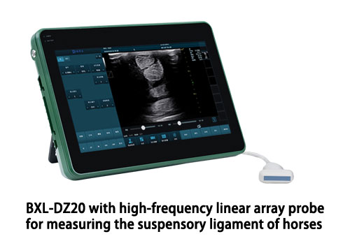
A high–frequency linear‐array transducer is essential for detailed imaging of the suspensory ligament. Linear probes in the 7.5–10 MHz range provide excellent resolution with sufficient depth penetration (4–6 cm) for most front and hind suspensory evaluations. While a standoff pad can improve probe‐to‐skin contact and image width, many practitioners omit it when using high‐frequency probes, opting instead to scan from both palmar and palmaro‐abaxial approaches to ensure full ligament coverage. Modern veterinary ultrasound systems also offer adjustable gain, focus, and dynamic range, which should be optimized to accentuate the fibrous architecture of the ligament.
Patient Handling and Site Preparation
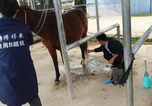
A calm, weight‐bearing stance is crucial for reproducible images. Sedation may be indicated for fractious horses, but should be used judiciously to avoid altering limb loading. Clip hair from the proximal suspensory origin (just distal to the carpus or hock) to the level of the fetlock. Clean the skin thoroughly with alcohol to remove dirt and debris, then apply generous ultrasound gel to eliminate air between the probe and skin. Position yourself to the side of the limb—facing the front limb or slightly behind the hind limb—to maintain safety and optimal probe orientation.
Scanning Protocol and Anatomical Landmarks
Systematically scan the suspensory ligament from its proximal origin to the branch insertions. Divide the metacarpal/metatarsal region into standard tiers—three equal sections between the carpometacarpal joint and the proximal extent of the palmar annular ligament—or use centimeter‐based landmarks referenced to the accessory carpal bone. Begin just distal to the carpus/hock, then move distally in 2–3 cm increments, acquiring both transverse and longitudinal images in each tier. Maintain consistent probe pressure and alignment to avoid anisotropy artifacts, which can mimic fiber disruption.

BXL-DZ20 Equine Suspensory Ligament Ultrasound
Transverse and Longitudinal Imaging
-
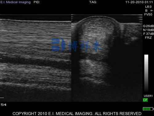
Transverse (cross‐sectional) views: With the probe perpendicular to the limb, the SL appears as an oval to triangular structure with homogeneous echogenicity. Carefully sweep medially and laterally to assess the full width of both the body and the branches.
-
Longitudinal (lengthwise) views: Rotate the probe 90° so that the beam runs parallel to the ligament fibers. Normal fibers appear as parallel, linear echogenic lines against a dark background. Pay special attention at the level of the proximal body where residual muscle fibers may be present, potentially masking pathology.
Identifying Suspensory Lesions
Ultrasonographic suspensory injuries are broadly categorized as:
-
Proximal Suspensory Desmitis (PSD) – enlargement and hypoechoic areas at the ligament origin just distal to the carpus or hock, commonly in hind limbs.
-
Body Lesions – focal fiber disruption in the mid‐body, seen as loss of parallel echogenic lines and localized hypoechoic defects.
-
Branch Injuries – swelling or fiber gaps in the medial or lateral branches near the sesamoid insertions.
Hypoechoic regions indicate acute fiber disruption or edema, while hyperechoic foci may represent mineralization or chronic scarring. Fluid surrounding the ligament suggests periligamentous inflammation or concurrent synovial involvement.
Measurement and Comparative Assessment
Quantitative assessment strengthens diagnostic confidence. Measure the cross‐sectional area (CSA) of the ligament at any lesion site and compare it to the contralateral limb; a >10–15% increase in CSA is considered significant in performance horses. Record echogenicity grades (e.g., normal, mildly hypoechoic, markedly hypoechoic) and fiber alignment (parallel, wavy, disrupted). Include distance markers or anatomical references on each image to ensure reproducibility in follow‐up exams.

Cross-sectional view of the suspensory ligament in the horse
Documentation and Follow‐Up
Save representative images from transverse and longitudinal planes at:
-
The origin (tier I)
-
Mid‐body (tier II)
-
Branch insertions (tier III)
-
Any abnormal areas
Include measurement overlays (CSA, width) and grading notes in the report. International practitioners recommend re‐examination every 30–60 days during rehabilitation to monitor healing trends in echogenicity and fiber realignment. Consistent image planes allow objective comparison over time.
Common Pitfalls and How to Avoid Them
-
Incomplete scanning: Failing to image all three regions can miss occult lesions.
-
Anisotropy artifacts: Incorrect probe angle can produce false hypoechoic bands; maintain the beam perpendicular to fibers.
-
Poor contact: Inadequate clipping or gel leads to image drop‐out; ensure full skin contact.
-
Misidentification of structures: The superficial digital flexor tendon and check ligaments lie adjacent to the SL—correlate anatomical landmarks meticulously.

Ultrasound image of the suspensory ligament in a horse
Advanced Modalities: Power Doppler and Elastography
Power Doppler ultrasonography can detect neovascularization associated with active desmopathy, offering adjunctive information to B‐mode imaging. Studies have shown increased Doppler signal in acutely injured suspensory branches compared to normal limbs, supporting its use in assessing tissue remodeling and inflammation. Elastography, though less widely available in veterinary practice, measures tissue stiffness and may help differentiate scar tissue from healthy ligament during rehabilitation.
Conclusion
Mastering the ultrasonographic examination of the equine suspensory ligament demands an understanding of its anatomy, meticulous technique, and consistent documentation. By combining high–frequency imaging, systematic scanning protocols, quantitative measurements, and advanced Doppler assessment, equine practitioners can accurately diagnose desmopathies, guide treatment, and monitor healing.
At BXL, we specialize in veterinary ultrasound solutions designed to meet the rigorous demands of musculoskeletal imaging in performance horses. Our high‐resolution systems and tailored transducer options empower veterinarians worldwide to “trust what they see” when evaluating soft tissue injuries.