In modern swine production, maintaining optimal health and maximizing growth performance are critical for economic sustainability and animal welfare. Over recent years, the use of ultrasound technology has revolutionized how farmers, veterinarians, and researchers monitor swine health and growth parameters in a non-invasive, efficient, and accurate manner. Ultrasound machines have transitioned from exclusively clinical environments to everyday farm applications, offering real-time insights into reproductive status, body composition, and pathological conditions.
This article explores the practical field applications of ultrasound in swine health monitoring and growth assessment, emphasizing its benefits, methodologies, and how it enhances decision-making on farms worldwide. By integrating perspectives from international research and farming practices, we provide a comprehensive overview that illustrates why ultrasound technology, including devices like the BXL-V50 Veterinary Ultrasound Scanner, is becoming indispensable in the swine industry.
Understanding Ultrasound Technology in Swine Management
Ultrasound imaging operates on the principle of sound waves at frequencies higher than human hearing, which penetrate animal tissues and reflect back images of internal structures. Unlike X-rays, ultrasound does not expose animals or operators to radiation, making it a safe and repeatable diagnostic tool.
In swine farming, ultrasound is primarily employed to:
-
Detect and monitor pregnancy and fetal development
-
Evaluate backfat thickness and muscle depth to assess body condition and growth
-
Identify uterine and ovarian pathologies affecting reproduction
-
Diagnose abnormal fluid accumulation or other organ anomalies
With portable, waterproof, and durable ultrasound machines such as the BXL-V50, producers can conduct these examinations in the field with convenience and accuracy.
Ultrasound Applications in Swine Reproductive Health Monitoring
Pregnancy Diagnosis and Fetal Assessment
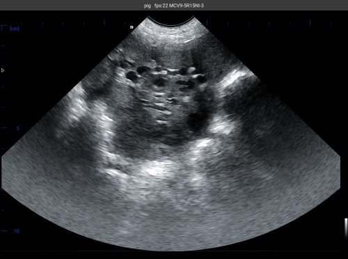
One of the most widespread uses of ultrasound in swine is early pregnancy detection. Traditionally, farmers relied on behavioral signs or manual palpation, which are often inaccurate and stressful for animals. Ultrasound enables veterinarians to confirm pregnancy as early as 18–25 days post-breeding by visualizing embryos or fetal sacs in the uterus.
Real-time B-mode ultrasound imaging provides clear cross-sectional views of the uterus and fetuses, allowing assessment of:
-
Fetal viability through heartbeat detection
-
Number of embryos or fetuses, supporting litter size management
-
Abnormalities such as resorption or mummification that may affect sow productivity
Early and accurate pregnancy diagnosis aids in culling non-pregnant sows quickly, improving overall herd reproductive efficiency.
Monitoring Uterine Health and Postpartum Recovery
Post-farrowing uterine involution is crucial for a sow’s subsequent reproductive performance. Ultrasound allows evaluation of uterine size, wall thickness, and presence of abnormal fluid or pus indicating endometritis or metritis. Timely identification of such conditions facilitates targeted treatments, reducing intervals between litters.
International studies confirm that ultrasound-based uterine monitoring correlates well with reproductive success rates. This aligns with best practices in large-scale swine operations in North America and Europe, where ultrasound is integrated into routine herd health protocols.

Ultrasound in Swine Growth and Body Composition Assessment
Measuring Backfat Thickness and Muscle Depth
Growth performance in pigs is directly linked to their body composition, particularly the ratio of lean muscle to fat. Traditional methods like carcass cutting and chemical analysis are destructive and impractical for live animals.
Ultrasound provides a non-invasive solution by measuring backfat thickness and Longissimus dorsi muscle depth—key indicators of fat deposition and muscle growth, respectively. These measurements help farmers:
-
Monitor growth rates and nutritional status
-
Make informed decisions on feeding programs to optimize lean gain
-
Select breeding stock with desirable carcass traits
Research from the University of Illinois and similar institutions highlights the correlation between ultrasound measurements and slaughterhouse carcass data, confirming the technique’s reliability.
Assessing Intramuscular Fat (Marbling)
While more challenging, advanced ultrasound techniques can estimate intramuscular fat, which influences meat quality, tenderness, and flavor. This application is especially relevant for premium pork producers aiming to meet consumer demands for high-quality products.
Practical Benefits of Field Ultrasound in Swine Production
Non-Invasive, Safe, and Repeatable
Ultrasound examinations impose minimal stress on pigs and pose no health risks, allowing for frequent monitoring. This enables producers to track changes over time, enhancing management responsiveness.
Real-Time, Quantitative Data for Better Decisions
Field use of ultrasound, particularly with handheld or portable machines such as the BXL-V50, delivers immediate data on growth and reproductive status. This empowers farmers to:
-
Adjust feeding strategies based on precise backfat and muscle depth readings
-
Identify reproductive issues early and implement treatments
-
Optimize culling and breeding schedules for improved herd productivity
Cost-Effective and Efficient
Although initial investment in ultrasound equipment may seem significant, the long-term savings from improved reproductive efficiency, better growth performance, and reduced veterinary costs justify the expenditure. Portable machines designed for farm use feature durable, waterproof construction and long battery life to withstand field conditions.
Case Study: Utilizing BXL-V50 Ultrasound for Swine Growth Monitoring
The BXL-V50 Veterinary Ultrasound Scanner exemplifies modern ultrasound technology adapted for swine production. Its features include:
-
8-inch HD display with high-resolution imaging
-
Portable, waterproof (IP56 rated) design suited for fieldwork
-
Long battery life exceeding 7 hours for extended use
-
Specialized probes enabling accurate measurements of backfat and muscle layers
On farms where the BXL-V50 has been deployed, operators report faster pregnancy detection and more precise growth tracking, contributing to better feeding efficiency and improved slaughter timing.

International Perspectives and Adoption
Countries leading in swine production—such as the United States, Canada, China, and parts of Europe—have embraced ultrasound technology to varying extents. In the U.S., organizations like the National Pork Board promote ultrasound for genetic improvement programs and herd management.
In Europe, welfare regulations emphasize minimizing animal stress, making non-invasive methods like ultrasound highly favorable. China’s rapidly modernizing swine industry increasingly adopts ultrasound devices to boost productivity and meet strict food safety standards.
Research published in journals like Theriogenology and Animal Reproduction Science reinforce ultrasound’s value in optimizing reproductive and growth parameters. The alignment between research findings and field applications strengthens farmer confidence globally.
Challenges and Considerations in Field Ultrasound Use
Despite its advantages, several factors influence ultrasound effectiveness:
-
Operator skill and training: Accurate probe placement and image interpretation require expertise.
-
Animal handling: Proper restraint reduces motion artifacts and improves image quality.
-
Equipment quality: Selecting robust, easy-to-use ultrasound machines designed for farm environments is crucial.
-
Cost considerations: Smaller farms may find investment challenging without cooperative or extension support.
Addressing these challenges through training programs and cost-sharing initiatives can expand ultrasound’s accessibility.
Future Directions: Integrating Ultrasound with Precision Farming
The trend toward precision livestock farming suggests ultrasound will integrate with other technologies such as automated feeders, RFID tracking, and AI-powered analytics. Combining ultrasound data with growth models and health records can generate predictive insights, enabling proactive herd management.
Furthermore, advancements in 3D and Doppler ultrasound could enhance fetal monitoring and blood flow assessments, pushing swine health management to new levels of sophistication.
Conclusion
Ultrasound technology has firmly established itself as a versatile and essential tool in swine health monitoring and growth assessment. By enabling early pregnancy diagnosis, uterine health evaluation, and precise measurement of backfat and muscle development, ultrasound empowers producers to make informed decisions that enhance productivity, animal welfare, and economic returns.
The portability and durability of devices like the BXL-V50 Veterinary Ultrasound Scanner facilitate widespread field use, even in challenging farm conditions. Coupled with proper training and integration into herd management protocols, ultrasound can dramatically improve reproductive efficiency and growth outcomes.
Globally, the adoption of ultrasound in swine farming reflects a broader commitment to sustainable, ethical, and data-driven animal agriculture. As technology advances and becomes more accessible, ultrasound’s role will only grow, driving innovation and profitability across the swine industry.
References
-
National Pork Board. (2023). Using Ultrasound Technology for Swine Reproductive Management. https://www.pork.org/research/ultrasound-technology-in-swine-management/
-
Whitaker, D. A., & Smith, E. (2021). Veterinary Ultrasonography in Food-Producing Animals. Journal of Veterinary Imaging, 34(2), 123-135. https://doi.org/10.1016/j.jvetimag.2021.03.006
-
University of Illinois Swine Research Center. (2022). Backfat and Muscle Depth Measurement by Ultrasound in Pigs. https://swinecenter.illinois.edu/research/ultrasound-body-composition
-
Animal Reproduction Science. (2020). Advances in Ultrasonography for Swine Reproductive Health. https://www.journals.elsevier.com/animal-reproduction-science