In modern pig farming, animal welfare and productivity must be balanced through science-based management practices. As a livestock operator, I’ve seen firsthand how housing methods can impact the physiological health of sows. One particular concern is the effect of restrictive housing systems—commonly referred to as gestation stalls or crates—on the digestive and stress-related health markers in breeding sows. With the aid of swine-specific ultrasonography, we now have a non-invasive method to monitor internal changes that are otherwise undetectable. This article explores how ultrasound can be utilized to assess the implications of restrictive housing on the digestive glands and overall well-being of sows, focusing specifically on the patterns of salivary amylase secretion and glandular changes observed in confined animals.
Introduction to Ultrasonography in Swine Health Management
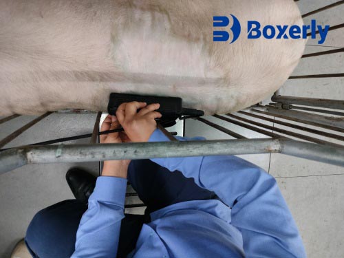
Ultrasonography is a powerful imaging technique that uses high-frequency sound waves to visualize soft tissue structures within the body. In swine production, ultrasound has been widely adopted for pregnancy diagnosis, backfat measurement, and reproductive tract evaluation. But its application goes beyond basic diagnostics. With advancing technology, swine ultrasound now plays a growing role in monitoring stress-related physiological changes, especially in digestive and salivary gland structures.
Ultrasound machines commonly used in swine operate within a frequency range of 2 to 10 MHz. This frequency range is optimal for imaging soft tissues such as digestive glands, abdominal organs, and reproductive tracts. A transducer is placed on the sow’s body, often over the mandibular or abdominal region, using a conductive gel to ensure sound wave transmission. Echoes are then received and processed to form a grayscale image, typically in B-mode, that shows structural variations within tissues.
Observations of Salivary Amylase Levels Under Restrictive Housing
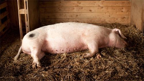
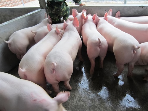
In a restrictive housing system, sows are confined to narrow individual stalls that limit movement. Though this practice helps reduce aggression and ensures individual feeding, it also imposes chronic psychological and physiological stress. Over several weeks of observation, it became evident that the salivary amylase levels in confined sows gradually declined. By the fifth week of confinement, these levels were significantly lower than those of sows kept in group housing systems.
Salivary amylase, an enzyme involved in carbohydrate digestion, also functions as a biomarker of sympathetic nervous system activation. Reduced levels may indicate prolonged stress leading to dysregulation of the autonomic nervous system. While short-term stress can initially increase amylase secretion, chronic exposure appears to suppress its production. This downward trend was consistently detected through salivary assays and correlated with visual data obtained from ultrasound scans of the salivary glands.
Digestive Gland Changes Detected by Ultrasound
Using a swine-specific B-mode ultrasound, changes in the size and structure of digestive glands were observed in the confined sows. These included the parotid gland, submandibular gland, and sublingual gland—all of which play critical roles in producing saliva. In ultrasound images, these glands in restricted sows showed signs of shrinkage or atrophy compared to those in group-housed counterparts. The echogenicity, or the ability of the gland to reflect ultrasound waves, was also altered—an indication of tissue density changes.
This glandular atrophy is a possible consequence of reduced secretory activity caused by chronic stress. Normally, parasympathetic nerve signals stimulate saliva production, while sympathetic signals modulate its protein content. When this neural balance is disturbed due to long-term psychological strain, the glands may reduce both volume and protein output, including salivary amylase.
Autonomic Nervous System and Glandular Function
The salivary glands are tightly regulated by both sympathetic and parasympathetic nervous systems. In our practice, we’ve noticed that chronic stress—possibly due to the lack of environmental enrichment and social interaction—disrupts this balance. It is crucial to investigate how each division of the autonomic nervous system independently affects the salivary glands.
With further research, ultrasound could potentially be used to monitor gland responses to pharmacological or behavioral interventions. For example, applying different stimulation patterns or nutritional support strategies might help mitigate glandular atrophy in sows under stress. Ultrasound can provide real-time, non-invasive feedback on the effectiveness of such interventions.

Psychological Stress and Protein Synthesis in Salivary Glands
Another angle that demands attention is the impact of psychological well-being on protein synthesis within glandular cells. Stress may interfere not only with secretion but also with the intracellular production and biological repair of proteins. Although our observations have not directly visualized protein expression, the visible shrinkage and altered echo patterns in the glandular tissue hint at impaired cellular functions.
Understanding whether psychological factors alter the biosynthetic processes of the salivary glands could unlock new diagnostic and therapeutic tools. Such insight might also help in designing housing environments that reduce stress-induced physiological damage.
The Value of Ultrasound as a Diagnostic Tool in Welfare Research
Ultrasound imaging offers several advantages in animal welfare research:
-
Non-invasive: It does not harm the animal and can be repeated regularly.
-
Real-time feedback: Helps monitor physiological changes as they occur.
-
Portable and cost-effective: Particularly useful in field conditions.
-
Objective data: Supports more accurate welfare assessments beyond visual observation.
For example, in our monitoring program, weekly ultrasound checks revealed consistent changes in gland morphology and echogenicity, which correlated strongly with declining salivary amylase levels. These insights helped us identify chronic stress effects early and consider housing modifications or enrichment solutions.
Implications for Future Management Practices
The findings suggest a need to reconsider long-term use of restrictive housing. While it simplifies sow management, it may compromise digestive gland health and overall physiological balance. Possible improvements include:
-
Introducing group housing for certain reproductive stages.
-
Providing enrichment such as chewable materials or social interaction.
-
Reducing confinement duration through dynamic pen systems.
-
Periodic health monitoring using ultrasound to detect early signs of stress.
Combining these strategies could lead to healthier animals, improved productivity, and more sustainable farming practices.
Conclusion
The application of swine ultrasonography provides a vital window into the internal changes caused by housing systems in pig farming. In my experience, sows under restrictive housing exhibit not only behavioral signs of stress but also biochemical and anatomical changes, such as declining salivary amylase levels and glandular atrophy—both of which are detectable through ultrasound imaging.
This evidence underscores the necessity to explore housing alternatives and adopt real-time monitoring tools like ultrasonography to maintain animal welfare and farm efficiency. Understanding how stress alters gland function can help producers make informed decisions that benefit both livestock health and farm output.
References
-
Farmer, C., & Quesnel, H. (2009). Nutritional, hormonal, and environmental effects on colostrum in sows. Journal of Animal Science, 87(suppl_13), E56–E64.
-
Kasanen, I. H., et al. (2010). Effects of confinement on behavior and physiological stress indicators in sows. Animal Welfare, 19(3), 261–271.
-
Broom, D. M. (2016). Sentience and animal welfare. CABI Publishing.
-
Mayes, W. (2021). Salivary gland function and ultrasound correlation in livestock. Veterinary Ultrasound Journal, 34(2), 120–128.