Yes, veterinarians can and do perform ultrasound examinations. In fact, ultrasonography has become a core diagnostic tool in modern veterinary practice, just like in human medicine. As a livestock farmer who works closely with veterinary professionals, I’ve personally witnessed how essential this imaging method is for diagnosing health issues in our animals — from cows and pigs to horses and sheep.
In this article, I will explain how veterinary ultrasound works, what it’s used for, and how it benefits both large and small animals on farms. I’ll write from the perspective of someone who relies on these services regularly and understands their practical value in everyday farm life.
Understanding Veterinary Ultrasound: A Farmer’s Perspective
Ultrasound, or ultrasonography, is likely the second most widely used imaging technique in veterinary clinics, after radiography (X-rays). But unlike X-rays, ultrasound doesn’t use harmful radiation. Instead, it uses high-frequency sound waves — typically between 1.5 and 18 MHz — to produce images of organs, tissues, and even blood flow within an animal’s body.
These sound waves are emitted by a handheld device called a transducer. When the transducer is placed on the animal’s body, it sends a burst of sound waves through the tissues. These waves bounce back at different speeds and intensities depending on the density of the structures they encounter. The returning echoes are captured by the transducer and translated by a computer into real-time images on a monitor.
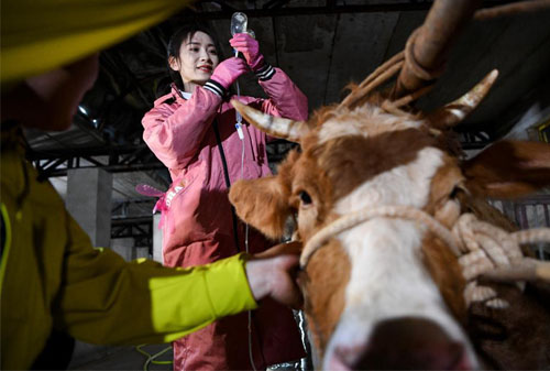
As someone managing a breeding operation, I’ve seen ultrasound used most frequently for reproductive checks — especially pregnancy diagnosis in cows and sows — as well as for evaluating soft tissue injuries and organ health in various animals.
How Vets Use Ultrasound on the Farm
On our farm, ultrasound is an essential tool during breeding seasons. For example, vets can perform transrectal ultrasounds on cows or mares to determine pregnancy just a few weeks after mating. This early confirmation helps us manage our breeding programs more efficiently, avoid wasted time, and reduce unnecessary costs from feeding non-pregnant animals.
Ultrasound is also frequently used to assess uterine health, ovarian follicles, and embryo viability. These checks are vital for maintaining herd fertility.
In pigs, ultrasound is used to measure backfat thickness — a key indicator of nutritional and reproductive status. The scan provides clear, reliable data without stressing the animals.
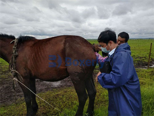
Beyond reproduction, veterinarians use ultrasound to examine the liver, kidneys, heart, bladder, intestines, tendons, and joints. For example, in horses, it’s the go-to method for detecting tendon or ligament injuries. In dogs or cats, it’s used to detect soft tissue lesions, monitor heart conditions, or even guide biopsies.
Real-Time Imaging: What Makes Ultrasound Special
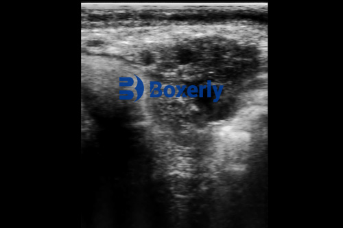
One thing that makes ultrasound so valuable is that it provides real-time imaging. The vet moves the transducer over the animal’s body, and the image updates instantly. This dynamic view helps in locating abnormalities, monitoring motion (such as a beating heart), or targeting specific organs.
The image can be frozen, saved, or even recorded as a short video. Most ultrasound machines follow the DICOM (Digital Imaging and Communications in Medicine) standard, ensuring high-quality, standardized image storage.
From a farmer’s perspective, this real-time functionality is a huge advantage. It means we get answers immediately, and vets can adjust their examination approach on the spot without delay.
Where Ultrasound Works Best — and Where It Doesn’t
Ultrasound is particularly good for evaluating soft tissues, like those in the abdomen, thorax, and reproductive tract. However, it doesn’t work well through air or bone. So, lungs and deeper bones are hard to image unless special techniques are used.
For instance, in cattle, bowel gas can sometimes block clear imaging of the surrounding abdominal organs. Similarly, in horses, bones may limit what can be seen beyond the joint surfaces.
Another limitation is depth. While some machines can scan up to 30 cm deep, image clarity can suffer at that depth because the returning sound waves are weaker. That’s why low-frequency transducers are used for deeper scans — although they sacrifice some image resolution.
Still, when the beam passes through fluid (like urine in the bladder), depth penetration improves, making it easier to see deeper organs. As farmers, we just need to understand that no tool is perfect, but ultrasound has far more benefits than drawbacks.
Special Applications: Echocardiography and Doppler Ultrasound
Ultrasound can also be used to evaluate the heart — a process called echocardiography. With M-mode and B-mode scans, veterinarians can see how heart valves and walls move. Advanced machines can even generate 3D images or calculate things like ejection fraction, cardiac output, and wall stiffness.
Doppler ultrasound adds another layer. It detects the speed and direction of blood flow by using the Doppler effect. This helps diagnose problems like valve insufficiency, stenosis, or abnormal blood circulation.
On our farm, echocardiography has been especially helpful in diagnosing older dogs or horses showing signs of fatigue or shortness of breath.
Ultrasound-Guided Biopsies: A Safer Option
Another practical use of ultrasound is in guiding biopsy instruments. Instead of blindly inserting a needle, the vet uses the ultrasound to pinpoint the exact location of a tumor or lesion. This is particularly useful for internal organs like the liver or kidneys.
This method not only increases accuracy but also reduces the need for open surgery. We’ve had animals that would have faced surgical risks otherwise — but thanks to ultrasound-guided biopsy, the diagnosis and treatment were much faster and safer.
Limitations and Interpretation Challenges
Even though ultrasound is sensitive, it’s not always specific. For example, pancreatitis in dogs may or may not show up clearly. Sometimes the pancreas is hard to access, or the changes are too subtle. Similarly, adrenal gland issues can be tricky to identify due to the presence of benign growths that may not cause symptoms.
This is where a veterinarian’s experience really matters. The echo patterns on the screen require skill and comparison with known healthy structures. For us farmers, this highlights the importance of having trained and experienced professionals perform the scan.

Conclusion: Why Ultrasound Matters in Veterinary Care
To answer the original question — yes, veterinarians can and absolutely do perform ultrasound exams. And from a livestock farmer’s perspective, I consider ultrasound an indispensable part of modern animal health care. It helps us make better decisions, detect problems early, and avoid unnecessary losses.
Whether it’s confirming pregnancy, assessing a tendon injury, or guiding a biopsy, ultrasound gives us powerful insights — all without surgery or radiation. As technology improves and vets gain more training, its value in both routine and emergency care will only continue to grow.