In modern cattle breeding, precision and timing are crucial to reproductive success. Artificial insemination (AI) is a widely adopted practice across the globe, yet its success hinges heavily on accurate heat detection, ovulation timing, and insemination technique. In recent years, siêu âm thú y—specifically B-mode ultrasonography—has emerged as a vital tool to support these aspects in cow reproduction. By providing real-time, non-invasive imaging of the reproductive tract, ultrasound enables veterinarians and technicians to optimize the timing and site of insemination and monitor uterine health before and after breeding. Trong bài viết này, I’ll share how veterinary ultrasound is transforming fertility outcomes in cows by improving insemination accuracy, supporting uterine health assessment, and guiding synchronized breeding programs.
Optimizing Insemination Site with Veterinary Ultrasound
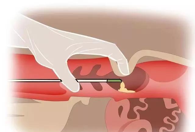
Understanding the cow’s reproductive anatomy is key to effective AI. Semen can be deposited in three primary locations: the cervix, the uterine body, and the uterine horns. Internationally, studies and field practices have shown that insemination directly into the cervix often leads to semen reflux, which lowers the number of viable sperm reaching the fertilization site. This is particularly problematic in high-value dairy herds, where reproductive efficiency is closely monitored.
In contrast, depositing semen too deeply—into the uterine horn—risks damaging the delicate endometrial lining, which can trigger inflammation and reduce conception rates. This makes the uterine body (specifically, its deeper segment) the ideal target site for AI, as it balances accessibility with minimal trauma to uterine tissues.
Veterinary ultrasound significantly enhances the accuracy of insemination by allowing real-time visualization of the uterine structure. With ultrasound guidance, technicians can confirm that the insemination pipette is positioned correctly within the uterine body, thereby reducing both under- and over-penetration. This technique has gained popularity in countries like the United States, the Netherlands, and Australia, where high-tech breeding systems are used to maximize conception rates.
The Role of Timing: Ovulation and Sperm Capacitation
Sperm deposited via AI must reach the oocyte in time to fertilize it, but neither too early nor too late. Ovulation timing varies based on breed, age, nutrition, climate, and hormonal synchronization protocols. Traditionally, AI timing relied on visual heat detection and manual rectal palpation to estimate ovarian activity. However, these methods are prone to error and subjectivity.
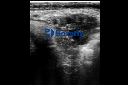
Veterinary ultrasound allows direct observation of ovarian follicles and the corpus luteum, giving a clear picture of the cow’s estrous stage. Through serial ultrasound examinations, technicians can track follicular growth and accurately predict the timing of ovulation. The optimal insemination window is typically 12 đến 18 hours after the onset of estrus, when the dominant follicle is near rupture and the uterus is most receptive.
Additionally, sperm capacitation—the biochemical transformation required for fertilization—takes several hours after insemination. Coordinating this process with ovulation is essential for success. By using ultrasound to time insemination precisely, reproductive specialists ensure that capacitated sperm are present in the oviduct when the egg is released.
Synchronized Breeding: The Power of Timed Insemination Programs
One of the most widely adopted innovations in global dairy and beef reproduction is estrus synchronization followed by timed artificial insemination (TAI). These protocols often involve hormonal treatments to synchronize ovulation across a group of cows, reducing the labor-intensive need to detect heat individually. However, success still depends on verifying that the cow’s reproductive tract is in a suitable condition.
Here, veterinary ultrasound again proves invaluable. By scanning the ovaries before TAI, technicians can ensure that a preovulatory follicle is present and that no abnormalities—such as cysts or an inactive corpus luteum—interfere with success. International research from universities such as Cornell (USA) and Utrecht (Netherlands) has confirmed that incorporating ultrasound into synchronization programs significantly boosts conception rates.
Assessing Uterine Health with Veterinary Ultrasound
Another major factor in successful fertilization is the health and readiness of the uterus. The uterus must be free of inflammation, Nhiễm trùng, or residual fluids from previous pregnancies to support sperm viability and embryo development. While cows have a natural ability to cleanse their uterine environment postpartum, this self-cleansing mechanism is not always sufficient.
Veterinary ultrasound provides a non-invasive way to assess uterine condition in detail. A healthy uterine lining appears uniform and echogenic on ultrasound, whereas signs of endometritis (uterine inflammation) or retained lochia can be easily identified as fluid pockets, thickened walls, or echogenic debris.
In countries with intensive dairy farming, such as New Zealand and Denmark, routine postpartum ultrasound checks are performed to ensure uterine involution is complete before cows enter the breeding cycle again. If issues are detected, targeted antibiotic or hormonal treatments can be administered, improving the odds of successful insemination at the next heat.
The Science Behind Uterine Self-Cleaning Mechanisms
Cows possess multiple physiological mechanisms to protect and purify their uterus after calving:
-
Uterine Contraction: Physical contractions help expel lochia and bacteria, which also reduce the availability of nutrients that could otherwise support microbial growth.
-
Immune Response: The endometrial surface is rich in immune cells, particularly neutrophils and macrophages. These cells engulf and digest pathogens through phagocytosis, much like in systemic infections.
-
Antibody Production: Local production of immunoglobulins helps neutralize toxins and prevent bacterial adherence to epithelial surfaces.
-
Uterine Secretions: Fluids in the uterus have natural antimicrobial properties, further promoting a sterile environment.
Ultrasound allows veterinarians to monitor the progression of these processes. If inflammation is seen resolving over time—indicated by the return of smooth, non-echoic endometrial layers—then the cow can safely reenter the reproductive cycle. On the other hand, persistent signs of infection may require intervention.
Practical Field Applications and Global Adoption
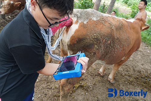
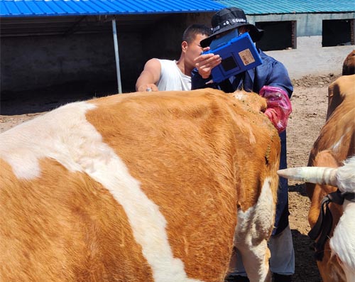
On commercial farms in the United States, Brazil, and parts of Europe, veterinarians now carry portable ultrasound machines during AI procedures. These compact, rugged devices offer real-time imaging and are often connected to mobile apps for data recording. This integration of imaging and data analytics allows better tracking of fertility trends, improving herd management.
From a practical perspective, this technology also helps reduce the variability introduced by different inseminators. In traditional rectal palpation-based AI, success depended heavily on the technician’s skill and experience. Ultrasound minimizes human error by providing visual confirmation of catheter placement, follicle development, and uterine health.
Challenges and Considerations
Despite its advantages, the use of veterinary ultrasound is not without challenges. Initial investment in ultrasound equipment can be high, and training is essential to interpret images accurately. In developing countries or smaller farms, access to skilled personnel and devices may still be limited. However, global trends indicate increasing affordability and availability of this technology.
Moreover, over-reliance on ultrasound without sound reproductive management principles—such as proper nutrition, hygiene, and cow comfort—will not guarantee results. Therefore, ultrasound should be viewed as one component of a comprehensive fertility program.
Kết thúc
Veterinary ultrasound has revolutionized artificial insemination in cows by enabling precision breeding practices. By optimizing insemination timing and site, monitoring uterine health, and supporting synchronization programs, this technology boosts conception rates and reduces reproductive losses. As ultrasound devices become more accessible and veterinarians become better trained, its adoption will likely continue growing across both developed and developing livestock systems.
For livestock producers committed to improving herd fertility, veterinary ultrasound offers not just a diagnostic tool, but a strategic advantage. Whether you manage a small family dairy in Ireland or a large beef operation in Argentina, integrating ultrasound into your reproductive protocols can lead to more pregnancies, healthier cows, and greater economic returns.