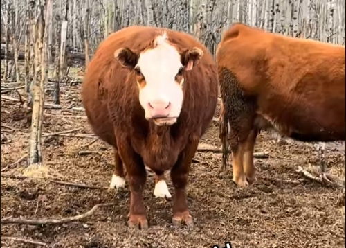
As a dairy producer, reproductive efficiency is the backbone of a successful herd. Timely ovulation in dairy cows is essential for synchronization protocols, conception rates, and overall milk production planning. However, delayed ovulation has become an increasingly observed issue across dairy farms worldwide. While the reasons vary—from poor nutrition to hormonal imbalances—siêu âm thú y has emerged as a vital diagnostic tool to identify, monitor, and manage delayed ovulation in dairy cattle.
Trong bài viết này, I’ll share how veterinary ultrasound is used to detect ovulatory problems in dairy cows, focusing on the underlying causes and what international studies have revealed about this challenge.
Understanding Ovulation in Dairy Cows
In a healthy reproductive cycle, the dominant follicle in a cow’s ovary matures under the influence of hormones and ovulates approximately 24 đến 30 hours after the preovulatory luteinizing hormone (LH) surge. Ovulation failure, or even a delay of several days, disrupts artificial insemination (AI) timing, leading to missed breeding windows and extended calving intervals.
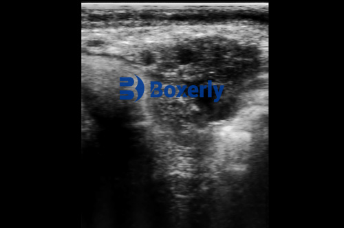
With the aid of real-time veterinary ultrasound, especially B-mode imaging, veterinarians and farm managers can track follicular development, monitor the timing of ovulation, and identify cows with delayed or absent ovulation. By repeatedly scanning the ovaries during estrus and following up post-insemination, it’s possible to determine whether ovulation occurred as expected or not.
Common Causes of Delayed Ovulation in Dairy Cows
-
Nutritional Imbalance and Poor Feed Management
One of the most frequently observed causes of delayed ovulation is improper nutrition—a topic well-documented in both Western literature and developing dairy regions.
⦁ Low-Quality Diets: When cows are underfed or fed low-energy, low-protein, or imbalanced rations, their body condition deteriorates. These cows may present with small, non-dominant follicles on ultrasound, indicating insufficient hormonal support to reach ovulation. Follicles often stagnate below 10–12 mm in diameter and fail to rupture.
⦁ Overfeeding and Obesity: On the flip side, over-conditioned cows often have excess adipose tissue that interferes with hormonal balance. Research from the University of Wisconsin suggests that obese cows experience altered insulin and leptin levels, leading to follicular cysts or anovulatory cycles. Ultrasound scans in such cows often reveal multiple fluid-filled follicles that remain static.
⦁ Unbalanced Rations: A deficiency in vitamins (especially A, D, and E) or trace minerals like selenium, copper, and zinc has been linked to poor follicular health. In North American herds, reproductive veterinarians report that ultrasound imaging shows underdeveloped follicles and low ovarian activity in herds fed diets lacking in micronutrient balance.
⦁ High Milk Output and Energy Deficits: In high-producing cows, nutrient demands for milk often exceed intake, especially in early lactation. Energy deficit reduces the availability of glucose and other metabolic substrates for the reproductive organs. This leads to impaired follicular development, as shown by reduced follicle number and growth rate in serial ultrasound observations.
-
Hormonal Imbalances
Hormones are the main regulators of ovulation, and any disruption in their synthesis, release, or timing can interfere with the process.
⦁ Insufficient LH Surge: LH is the key trigger for ovulation. If the hypothalamus or pituitary fails to release a timely and adequate LH surge, ovulation does not occur. On ultrasound, these cows often show follicles that persist for multiple days without rupture, sometimes transitioning into cystic structures.
⦁ Elevated Prolactin (PRL): Research from Dutch and Canadian veterinary colleges suggests that high-yielding cows may have elevated PRL levels postpartum. PRL can interfere with LH receptor sensitivity on follicles, inhibiting the final maturation process. Ultrasound studies show multiple small follicles with minimal growth and no signs of ovulation in these animals.
⦁ Prostaglandin (PG) Deficiency: PGs are crucial in breaking down follicular walls during ovulation. A shortage of PG—especially PGF2α—can delay follicle rupture. In affected cows, the follicles grow to near-ovulatory size but remain intact on ultrasound, even days past estrus.
⦁ Estrogen-androgen Imbalance: A reduction in estrogen production and a concurrent rise in intra-follicular androgens can cause follicular atresia or “follicle lock.” Using ultrasound, such follicles appear irregular in shape, with echogenic contents suggestive of degenerative changes.
⦁ Reduced Enzyme Activity: Ovulation is not purely hormonal—it also involves the action of proteolytic enzymes such as collagenase and plasminogen activators that weaken the follicular wall. When enzyme activity is reduced, either genetically or due to nutritional stress, the follicle cannot rupture. Veterinary ultrasound reveals mature-looking follicles that remain unruptured for several estrous cycles.
-
Environmental and Management Factors
⦁ Lack of Physical Activity: Exercise influences blood circulation and hormone metabolism. In confined dairy barns with limited space, cows may lack sufficient exercise, leading to suppressed ovarian activity. On ultrasound, this often translates to a lack of dominant follicles or slow follicular turnover.
⦁ Heat Stress: In warmer climates, heat stress significantly impairs reproduction. According to research from Israel and Florida, high ambient temperatures reduce estradiol secretion, alter LH pulse frequency, and delay ovulation. Ultrasound tracking during summer months shows extended follicular phases and poor ovulatory rates.
⦁ Improper Estrus Detection and Synchronization Protocols: Mistimed artificial insemination due to poor estrus detection or misapplication of synchronization hormones can make it appear as though ovulation is delayed when, in fact, timing was simply off. However, ultrasound can confirm true ovulation by scanning 24–36 hours post-AI and verifying corpus luteum (CL) formation.
Role of Veterinary Ultrasound in Diagnosis and Prevention
Veterinary ultrasound allows for detailed, non-invasive, and repeatable observation of the ovarian structures. When diagnosing delayed ovulation, we typically look for:
-
Follicular diameter and shape
-
Wall thickness and echogenicity
-
Presence or absence of CL (indicating successful ovulation)
-
Rate of follicular turnover across cycles
By performing routine reproductive ultrasound exams, we can identify cows at risk for delayed ovulation early and take action—whether that means adjusting diets, correcting hormonal treatments, or modifying herd management strategies.
Best Practices Adopted by International Farms
Progressive dairy farms in the U.S., Europe, and New Zealand integrate ultrasound with hormonal protocols to increase reproductive efficiency. They often:
-
Scan all cows post-insemination at day 10–14 to confirm ovulation
-
Use ultrasound to customize synchronization programs based on individual ovarian response
-
Monitor follicular growth during superovulation for embryo transfer programs
-
Record ultrasound metrics to inform veterinary reproductive consultation
Kết thúc
Delayed ovulation is a complex issue with multifactorial origins—nutrition, hormones, and management all play a role. Fortunately, veterinary ultrasound provides a clear window into the cow’s reproductive system, allowing veterinarians and producers to detect, understand, and respond to ovulatory issues in real-time.
As global dairy systems increasingly shift toward precision livestock farming, incorporating ultrasonography into routine herd health protocols will be vital for sustaining fertility, productivity, and profitability.