For modern dairy farmers, managing reproductive health is essential to maintaining productivity and profitability. Among the many reproductive disorders that threaten high-yield dairy herds, ovarian cysts stand out as a particularly challenging problem. These cysts disrupt normal estrous cycles, reduce conception rates, and contribute to prolonged calving intervals—all of which reduce milk output and economic returns. Fortunately, veterinary ultrasonography has become an invaluable tool for the early detection and management of ovarian cysts. În acest articol, I will explore how animal ultrasound scanners are used to identify ovarian cysts in high-yield dairy herds, share insights from my own experience, and highlight why this technology is increasingly adopted by farmers worldwide.
Understanding Ovarian Cysts in Dairy Cattle
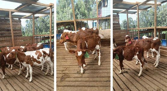
In high-producing dairy cows, ovarian cysts are a common reproductive pathology, especially during the early postpartum period when metabolic demands are intense. An ovarian cyst is typically defined as a fluid-filled structure greater than 25 mm in diameter that persists for more than 10 days without the presence of a corpus luteum (CL). There are generally two types of ovarian cysts: follicular cysts and luteal cysts.
-
Follicular cysts are thin-walled, estrogen-secreting, and prevent normal ovulation.
-
Luteal cysts have a thicker wall and secrete progesterone, mimicking a persistent CL and preventing normal estrus behavior.
Ovarian cysts disrupt reproductive cycles, leading to silent heat, irregular estrus behavior, or complete anestrus. For high-yield dairy herds that depend on timely calving to sustain milk production, the economic impact of undiagnosed or mismanaged cysts can be significant.
The Role of Ultrasonography in Ovarian Cyst Diagnosis
In the past, diagnosing ovarian cysts relied heavily on rectal palpation, a skill-dependent and somewhat subjective method. Today, veterinary ultrasonography offers a more precise, non-invasive, and reliable alternative. The real-time imaging capabilities of an animal ultrasound scanner allow veterinarians and trained farm technicians to distinguish between normal ovarian structures and pathological cysts with high accuracy.
As a farmer, I have found that using an ultrasound scanner—particularly a high-resolution B-mode machine—dramatically improves the early detection of ovarian cysts. Unlike palpation, ultrasound allows me to directly visualize the ovaries, measure the size of the structures, assess the wall thickness, and even evaluate internal echoes that can differentiate follicular from luteal cysts.
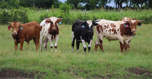
Why High-Yield Dairy Cows Are More Prone to Ovarian Cysts
Numerous scientific studies from around the world have shown that high-yield dairy cows are at greater risk for ovarian cyst development due to the metabolic stress associated with heavy lactation. In countries like the United States, Canada, and New Zealand, where dairy cows routinely produce upwards of 10,000 liters of milk per lactation, metabolic imbalances—particularly negative energy balance (NEB)—are common during early lactation. This metabolic stress disrupts the normal secretion of gonadotropin-releasing hormone (GnRH) and luteinizing hormone (LH), both critical for ovulation.
Suplimentar, the selection for high milk yield has inadvertently led to cows with longer postpartum anestrous periods, making them more vulnerable to cyst formation. Foreign research (Lucy, 2001; Dobson et al., 2007) consistently emphasizes the link between intensive milk production and reproductive dysfunction.
How We Use Ultrasound to Detect Ovarian Cysts on Our Farm
On my farm, routine ultrasound examinations are integrated into our reproductive management program. Using a portable veterinary ultrasound scanner, I begin scanning cows as early as 30 days postpartum. The scanning process is straightforward:
-
Transrectal Approach: The probe is gently inserted into the rectum while the cow is properly restrained.
-
Ovary Identification: The ovaries are located by sweeping the probe laterally from the uterine horns.
-
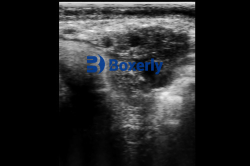
Image Interpretation: Real-time B-mode images display the ovarian structures. Cysts appear as large, circular, fluid-filled anechoic (black) areas. Wall thickness and internal contents provide critical information.
-
Measurement: Using on-screen calipers, I measure cyst diameter and wall thickness. Follicular cysts typically have thin walls (<3 mm), while luteal cysts display thicker walls (>3 mm) and may have internal echoes.
By systematically recording these measurements, I can track the progression or regression of cysts over time and adjust treatment protocols accordingly.
Key Advantages of Ultrasound Over Traditional Methods
In my experience, the shift from palpation to ultrasound scanning offers several major advantages that are particularly relevant for high-yield dairy herds:
-
Accuracy and Objectivity: Ultrasound provides clear visual confirmation of cyst presence, size, and type. This reduces reliance on subjective palpation skills.
-
Early Detection: Scanning allows for the detection of cysts before clinical symptoms appear, improving treatment outcomes.
-
Differentiation: The ability to distinguish between follicular and luteal cysts helps determine the most appropriate hormonal treatment (De ex., GnRH for follicular cysts; PGF2α for luteal cysts).
-
Non-invasive and Low-Stress: Ultrasound exams are quick and cause minimal discomfort, reducing stress in already metabolically challenged cows.
-
Monitoring Treatment Response: Repeat scans help assess whether treatment protocols are effective or require adjustment.
Ultrasound as a Preventive Reproductive Management Tool
Beyond simply diagnosing ovarian cysts, ultrasound scanning plays a crucial preventive role on our farm. We schedule routine reproductive ultrasound exams for all cows at key stages:
-
Postpartum Health Checks (30-45 Days): To detect any early reproductive disorders.
-
Pre-Breeding Exams (60 Days): To ensure cows are cycling normally before insemination.
-
Pregnancy Confirmation (30-60 Days Post AI): To verify successful conception and identify early embryonic loss.
This comprehensive approach has reduced the incidence of open cows, shortened calving intervals, and improved overall herd reproductive efficiency. Research from Europe (Peter et al., 2009; López-Gatius et al., 2010) supports this strategy, showing that herds with routine ultrasound programs consistently outperform those relying solely on palpation.
Case Example: Ultrasound Makes a Difference
One of our top-producing cows, a Holstein named Daisy, was not coming into heat as expected 90 days postpartum. Palpation suggested a possible cyst, but we confirmed it with ultrasound: a 32 mm fluid-filled structure with a thin wall—a classic follicular cyst.
Based on the ultrasound findings, we administered GnRH, followed by PGF2α a week later. A follow-up scan 10 days later showed the cyst had resolved, și Daisy displayed clear estrus behavior. She was successfully inseminated and later confirmed pregnant. Without ultrasound, we may have either delayed treatment or administered inappropriate hormones.
Advances in Portable Ultrasound Technology
Modern Ecografie veterinară devices have become increasingly portable, ușor de utilizat, and affordable. On our farm, we use a model equipped with an 8-inch HD display, waterproof casing, and long battery life, making it ideal for field conditions. Devices like the BXL-V50 provide excellent image clarity even under challenging farm environments, allowing both veterinarians and trained farm staff to conduct accurate scans.
The affordability and ease of training associated with portable ultrasound units have made this technology widely accessible, not only on large commercial farms in North America and Europe but also increasingly in developing dairy industries in Asia, South America, and Africa.
Limitations and Challenges
While ultrasound is a powerful diagnostic tool, it does have some limitations:
-
Training Required: Operators need proper training to accurately interpret images, particularly in distinguishing subtle differences between cyst types.
-
Equipment Cost: Although prices have dropped, high-quality machines still represent a significant investment for smaller farms.
-
Scheduling and Labor: Incorporating routine scanning requires organization and labor allocation, especially on large farms with hundreds of cows.
Însă, the long-term benefits of improved reproductive efficiency and reduced economic losses easily justify these challenges.
Global Perspectives: Ultrasound Adoption Across Countries
Internationally, the adoption of ultrasonography for ovarian cyst diagnosis is rapidly expanding. De exemplu:
-
In the United States, ultrasound is now standard practice on most large-scale dairies. The National Animal Health Monitoring System (NAHMS, 2018) reports over 70% of herds use some form of ultrasound for reproductive management.
-
In Canada, research from the University of Guelph emphasizes its role in managing reproductive disorders in high-yielding Holsteins.
-
In Europe, routine ultrasonography is often incorporated into intensive reproductive programs, especially under strict animal welfare and productivity regulations.
-
In New Zealand, pasture-based dairy systems still rely heavily on seasonal breeding, but ultrasound is increasingly used during fixed-time artificial insemination programs to optimize conception rates.
This global trend reflects growing recognition that precision reproductive management is critical for sustaining profitability in modern dairy systems.
Economic Impact of Ultrasound Use in High-Yield Herds
Studies have consistently shown that incorporating ultrasound into herd management leads to significant economic gains. These benefits include:
-
Reduced days open
-
Higher conception rates
-
Fewer culled cows due to reproductive failure
-
Optimized hormone use, reducing drug costs
-
Higher lifetime milk yield per cow
Un 2021 study published in the Journal of Dairy Science (Giordano et al.) estimated that effective reproductive monitoring with ultrasound can improve net profits by $100–$300 per cow annually in high-producing herds.
Concluzie
For today’s dairy producer, maintaining reproductive efficiency is one of the most controllable levers for maximizing profitability. Ovarian cysts, while common, are no longer as intimidating thanks to the widespread availability of veterinary ultrasound technology. By integrating regular ultrasound scanning into herd management, farmers like myself can detect ovarian cysts early, apply appropriate treatments, and ultimately reduce reproductive losses.
As global demand for milk continues to rise, high-yield dairy herds must operate with ever-greater precision. Veterinary ultrasound scanners offer a real-time, non-invasive, and highly accurate window into reproductive health. On my farm, this technology has become indispensable—not only for identifying ovarian cysts but also for optimizing every stage of the reproductive cycle.

With portable, high-definition machines like the BXL-V50, even smaller farms can now access cutting-edge diagnostic capabilities once reserved for large commercial operations. As more farmers worldwide adopt this approach, the future of dairy farming looks increasingly data-driven, efficient, and sustainable.
Reference Sources:
-
Lucy, M. C. (2001). Reproductive loss in high-producing dairy cattle: where will it end? Journal of Dairy Science, 84(6), 1277-1293. https://doi.org/10.3168/jds.S0022-0302(01)70158-0
-
Dobson, H., et al. (2007). The high-producing dairy cow and its reproductive performance. Reproduction in Domestic Animals, 42, 17-23. https://doi.org/10.1111/j.1439-0531.2007.01014.x
-
Peter, Un. T., et al. (2009). Use of ultrasonography in cattle reproduction. Theriogenology, 71(6), 977-984. https://doi.org/10.1016/j.theriogenology.2008.11.011
-
López-Gatius, F. (2010). Is fertility declining in dairy cattle? A retrospective study in northeastern Spain. Theriogenology, 73(5), 637-644. https://doi.org/10.1016/j.theriogenology.2009.10.001
-
Giordano, J. O., et al. (2021). Economic evaluation of reproductive programs using ultrasound in dairy cows. Journal of Dairy Science, 104(3), 2931-2947. https://doi.org/10.3168/jds.2020-19490
-
National Animal Health Monitoring System (NAHMS). (2018). Dairy Cattle Management Practices in the United States. USDA.