Embryo transfer (ET) has become a transformative reproductive biotechnology widely used in livestock breeding to accelerate genetic improvement, increase reproductive efficiency, and preserve valuable genetics. Central to the success of ET programs is the precise and minimally invasive retrieval of oocytes (egg cells) from donor females. Veterinary ultrasound technology plays a pivotal role in visualizing and guiding this oocyte retrieval process, enhancing accuracy, reducing complications, and improving overall outcomes.
În acest articol, I will explore how veterinary ultrasound technology is applied in oocyte retrieval during embryo transfer procedures, discuss the technical nuances, and explain why this imaging method is indispensable in modern animal reproduction management worldwide. Drawing from international practices and scientific studies, this comprehensive review aims to highlight the advances and benefits of ultrasound-guided oocyte retrieval in the livestock industry.
The Role of Oocyte Retrieval in Embryo Transfer Programs
Embryo transfer involves collecting embryos from genetically superior donor females and transferring them to recipient females who will carry the pregnancy to term. The process begins with stimulating the donor’s ovaries to produce multiple follicles, from which oocytes are retrieved. These oocytes are either fertilized in vitro (in the laboratory) to produce embryos or used directly for in vivo fertilization and embryo collection.
The efficiency of embryo transfer programs hinges on several factors, among which the successful retrieval of high-quality oocytes is fundamental. Traditional oocyte retrieval methods were often blind or relied on palpation techniques, leading to variable success rates and increased animal stress or injury. Veterinary ultrasound technology has revolutionized this step by providing real-time visualization of ovarian structures, enabling precise follicular puncture and oocyte aspiration.
Veterinary Ultrasound Technology in Oocyte Retrieval: An Overview
Ultrasound uses high-frequency sound waves to create live images of internal tissues. În medicină veterinară, ultrasound devices adapted for reproductive applications enable clinicians to observe ovarian follicles, measure follicle size, and guide fine needles during oocyte aspiration.
Two main ultrasound modalities are applied in oocyte retrieval:
-
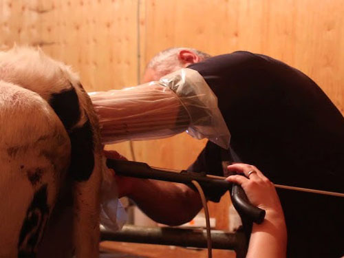
Transrectal ultrasound: Commonly used in cattle, where the probe is inserted into the rectum, allowing close proximity to the ovaries for high-resolution imaging.
-
Transvaginal ultrasound: More frequent in small ruminants or equine species, involving a vaginal probe for imaging ovaries and guiding needle placement.
Regardless of the species, veterinary ultrasound provides critical real-time feedback during oocyte retrieval, ensuring accuracy and minimizing trauma.
Imaging Ovarian Follicles and Identifying Target Oocytes
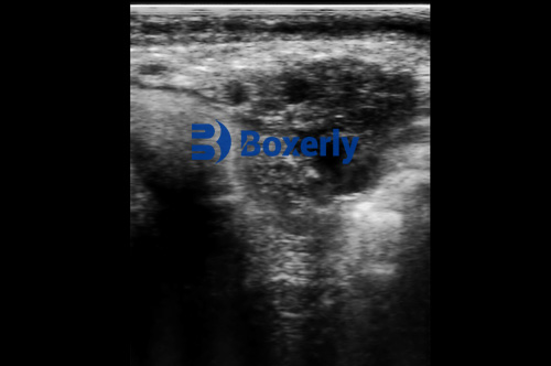
The ultrasound screen displays follicles as anechoic (dark) circular structures within the ovarian stroma. Follicle size is a key indicator of oocyte maturity, and ultrasound enables precise measurement of these follicles, typically ranging from 6 spre 20 millimeters depending on species and reproductive stage.
Identifying the right follicle is essential because immature follicles yield oocytes with low developmental potential, while overly large follicles may have already ovulated or contain aged oocytes. Ultrasound allows veterinary technicians and veterinarians to select follicles with optimal characteristics, increasing the likelihood of successful fertilization and embryo development.
Guiding Needle Aspiration
During oocyte retrieval, a specialized needle is inserted through the vaginal or rectal wall into the follicle under ultrasound guidance. The real-time imaging helps confirm the needle’s position within the follicle and monitor follicular fluid aspiration.
The use of Doppler ultrasound can further aid by visualizing blood flow around the ovary, helping avoid vascular damage and reducing the risk of hemorrhage. The combination of 2D imaging and Doppler technology enhances safety and improves the precision of oocyte recovery.
Advantages of Ultrasound-Guided Oocyte Retrieval in Embryo Transfer
Worldwide, veterinary practitioners and livestock producers have embraced ultrasound technology in oocyte retrieval because it offers multiple significant benefits:
Non-Invasive and Reduced Animal Stress
Traditional methods like laparotomy (surgical opening of the abdomen) or laparoscopic retrieval, though effective, are invasive and require anesthesia with prolonged recovery. Ultrasound-guided transvaginal or transrectal oocyte retrieval is minimally invasive, often performed with sedation or light restraint. This reduces animal discomfort, lowers infection risk, and allows quicker return to normal activity.
Improved Retrieval Rates and Oocyte Quality
By enabling direct visualization, ultrasound guidance improves the retrieval rate of viable oocytes per donor session. Studies have demonstrated that ultrasound-guided aspiration yields higher-quality oocytes with better maturation status, translating into improved fertilization rates and embryo viability.
Real-Time Feedback and Procedural Control
Ultrasound imaging gives the operator immediate feedback on needle placement and follicular aspiration success. This reduces guesswork and procedural errors, improving overall efficiency and reducing procedure time.
Cost-Effectiveness and Practicality
Though ultrasound equipment requires upfront investment, the increased efficiency and reduced animal health complications lead to cost savings. Portable veterinary ultrasound machines allow on-farm procedures, decreasing the need to transport valuable donors and improving logistical convenience.
Technical Considerations in Ultrasound Visualization of Oocyte Retrieval
While the benefits are clear, effective ultrasound-guided oocyte retrieval requires technical expertise and equipment optimization.
Probe Selection and Frequency
Higher-frequency probes (7.5 MHz and above) offer superior resolution for imaging small follicles but have limited tissue penetration. Lower-frequency probes penetrate deeper but with lower image clarity. Selecting the right probe depends on species, ovarian size, and access route.
Needle Design and Visualization
The needle used for aspiration must be compatible with ultrasound imaging. Echogenic needles, designed with textured surfaces or coatings to enhance visibility on ultrasound, help operators track needle advancement and placement precisely.
Operator Training
Competent use of ultrasound technology demands training in reproductive anatomy, ultrasound physics, and hand-eye coordination for needle manipulation. Many veterinary training programs now include specific modules on ultrasound-guided reproductive techniques.
Environmental and Animal Factors
Optimal imaging requires proper animal preparation, inclusiv restraining techniques and sometimes bowel emptying in cattle to reduce interference. Ambient lighting and machine settings (gain, depth, focus) must be adjusted to optimize image clarity.
International Perspectives and Applications in Livestock Breeding
Globally, countries with advanced livestock industries have adopted ultrasound-guided oocyte retrieval as standard practice in embryo transfer. De exemplu:
-
Statele Unite: The dairy and beef cattle sectors utilize transrectal ultrasound to retrieve oocytes in superovulated donors for embryo transfer and in vitro fertilization programs.
-
Europe: Small ruminant industries in France and Spain apply transvaginal ultrasound in sheep and goats, increasing genetic gain in fine wool and meat production breeds.
-
Australia and New Zealand: Emphasizing animal welfare, veterinary ultrasound-guided retrieval is widespread in cattle and equine reproduction centers, coupled with emerging in vitro embryo technologies.
-
Asia: Rapid growth in pig and cattle industries has driven ultrasound adoption, especially for genetic improvement in high-value breeds.
These international experiences underscore ultrasound’s pivotal role in improving reproductive eficiență, animal welfare, and genetic progress in various production systems.
Case Studies Demonstrating Ultrasound-Guided Oocyte Retrieval Success
Case Study 1: Dairy Cattle in the USA
A commercial dairy farm implementing ultrasound-guided transrectal oocyte retrieval reported a 25% increase in embryo yield per donor compared to conventional palpation methods. The precise follicle targeting reduced the average procedure time and lowered donor stress indicators measured by cortisol levels.
Case Study 2: Sheep Breeding in Spain
A research facility in Spain employed transvaginal ultrasound for oocyte retrieval in a fine wool sheep breed. The study documented that ultrasound guidance increased viable oocyte recovery by 30%, facilitating accelerated genetic selection for fleece quality.
Case Study 3: Equine Embryo Transfer in Australia
Australian equine reproduction centers using ultrasound-guided follicular aspiration for in vitro fertilization observed improved fertilization rates and embryo viability, contributing to more efficient breeding programs for elite sport horses.
Challenges and Future Directions in Ultrasound Visualization of Oocyte Retrieval
While ultrasound technology has transformed oocyte retrieval, some challenges remain:
-
Equipment Cost and Access: High-quality ultrasound machines and compatible aspiration needles can be costly, limiting use in low-resource settings.
-
Learning Curve: Mastery of the technique requires training and practice, which may slow adoption.
-
Imaging Limitations: In large animals with deep ovaries or excessive fat, imaging clarity may be reduced.
Future developments include:
-
Enhanced Needle Visualization: New echogenic materials and needle designs will improve operator confidence.
-
3D and 4D Ultrasound: Advanced imaging modalities promise more detailed visualization of follicle and oocyte morphology.
-
Integration with Robotic Assistance: Automation of needle guidance could further reduce procedure variability and improve outcomes.
-
Ultrasunete portabile Innovations: Mici, wireless ultrasound units will increase accessibility for remote or on-farm applications.
Concluzie
Veterinary ultrasound technology has become an indispensable tool for visualizing and guiding oocyte retrieval processes in embryo transfer procedures. By enabling real-time, accurate imaging of ovarian follicles and precise needle placement, ultrasound enhances retrieval success, improves animal welfare, and drives genetic advancement in livestock industries worldwide.
As ultrasound equipment becomes more affordable and training more widespread, its application in oocyte retrieval is poised to expand further. The benefits—ranging from increased embryo yield and better reproductive efficiency to reduced animal stress—highlight the transformative impact of ultrasound tech in reproductive management.
For livestock farmers, veterinarians, and reproductive specialists, mastering ultrasound-guided oocyte retrieval is not only a technical skill but a strategic advantage in achieving sustainable and profitable breeding programs.
References
-
Hasler, J. F. (2014). In vitro embryo production and transfer in cattle. Theriogenology, 81(1), 102-111. https://doi.org/10.1016/j.theriogenology.2013.09.022
-
Merton, J. S., Hasler, J. F., & Schaeffer, L. R. (2018). Assisted reproductive technologies in livestock production. Reproduction in Domestic Animals, 53(Suppl 2), 17-28. https://doi.org/10.1111/rda.13292
-
Pugliesi, G., & Hennebold, J. D. (2019). Advances in imaging ovarian follicle dynamics: applications to oocyte retrieval. Frontiers in Veterinary Science, 6, 85. https://doi.org/10.3389/fvets.2019.00085
-
Society for Theriogenology. (2023). Ultrasound guidance for oocyte retrieval in cattle. https://www.therio.org/ultrasound-guidance-oocyte-retrieval