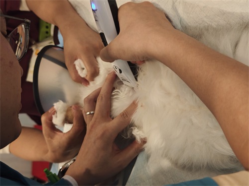
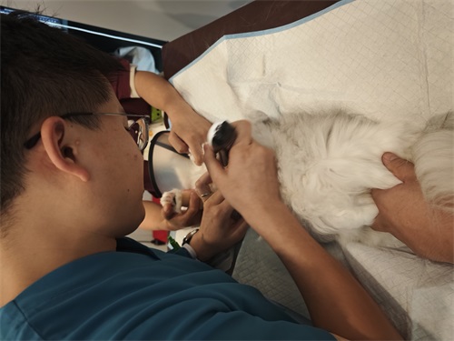
For breeders, veterinarians, and pet owners, successful canine reproduction hinges on timing, precision, and monitoring. Modern veterinary medicine increasingly relies on advanced imaging tools to improve reproductive success in dogs. Among these, ultrasound—especially B-mode ultrasonography—has emerged as a highly reliable, non-invasive, and informative tool. It allows the visualization of key reproductive structures, monitoring of follicular development, pregnancy diagnosis, Sănătatea fetală, and even disease detection. This article explores how veterinary ultrasound is applied across different stages of the canine reproductive cycle, referencing both clinical experience and global veterinary practices.

Monitoring Ovarian Development for Optimal Breeding
One of the most important applications of ultrasound in canine reproduction is monitoring the ovaries to determine the ideal breeding time. A female dog’s ovary is relatively small, typically measuring 1.5–3.0 cm × 0.7–1.5 cm × 0.5–0.75 cm. Because of its small size and the surrounding fat tissue, ultrasound images can be obscured. Skilled probe placement and scanning technique are essential for accurate visualization.
Using ultrasonography, the ovaries—often described as mulberry-shaped—can be located near the third or fourth lumbar vertebra, approximately 1–4 cm caudal to the kidneys. In bitches that have already given birth, the ovaries tend to be positioned further back and lower in the abdomen. During estrus, typically around 2–3 days post-onset, developing follicles appear as multiple circular anechoic (black) regions on ultrasound, sometimes reaching a diameter of up to 13 mm.
Understanding the progression of follicular development is crucial. In proestrus and early estrus (days 1–8), follicles grow slowly. From day 10 spre 15, they expand rapidly and then stop growing—signaling that ovulation is near. This phase is the ideal window for natural mating or artificial insemination. Ultrasound not only confirms the presence of mature follicles (>3 mm in diameter) but also helps count them across different planes.
When only a few mature follicles are observed or their development is asynchronous, gonadotropin treatments like FSH, PMS-G, or hCG can be administered to stimulate ovulation. This approach, backed by ultrasound evidence, improves conception rates and litter sizes.

Observing Embryonic Development and Monitoring Pregnancy
Ultrasound plays a central role in confirming pregnancy, estimating fetal age, counting fetuses, and detecting pregnancy-related complications. Starting from 15 spre 20 days post-mating, pregnancy sacs (gestational sacs) can be identified on ultrasound. Around days 23–25, the embryo becomes visible as a bright echoic structure within the chorionic cavity. By days 21–28, the fetal heartbeat can be detected—a reliable sign of pregnancy viability.
As pregnancy progresses, more anatomical details become visible:
-
Days 26–28: Clear visualization of head and trunk
-
Days 31–35: Differentiation of limbs and choroid plexus
-
Days 35–40: Beginning of fetal bone mineralization
-
Days 40–47: Identification of optic vesicles and internal organs
Using these anatomical landmarks, veterinarians can estimate fetal age with reasonable accuracy. Fetal count can be approximated based on the number of gestational sacs and embryos observed—though this estimate becomes less reliable in late gestation due to overlapping fetuses.
Ultrasound also facilitates early identification of reproductive complications. De exemplu, a lack of fetal heartbeat indicates embryonic or fetal death. Echoic gas bubbles suggest fetal maceration, while reduced sac size may indicate embryo absorption. Close monitoring of uterine content during and after delivery helps assess whether parturition has concluded and whether the uterus is involuting properly.
Diagnosing Reproductive Disorders
Beyond its utility in breeding and pregnancy, ultrasound is an essential diagnostic tool for a wide range of reproductive disorders in dogs. Common conditions include:
-
Pyometra and hydrometra: Ultrasound reveals fluid-filled uterine horns, distinguishing between infection and sterile fluid accumulation.
-
Ovarian cysts: These appear as round, anechoic or hypoechoic structures on the ovaries and can disrupt normal estrous cycling.
-
Dystocia and miscarriage: Structural abnormalities or retained fetal parts can be seen, aiding in deciding the necessity for surgical intervention.
-
Tumors of the uterus, mammary glands, and testicles: Ultrasound offers insight into size, location, and composition (solid vs. cystic) of abnormal growths.
-
Prostatic diseases: In male dogs, benign prostatic hyperplasia, prostatitis, and neoplasia are common. Ultrasound helps differentiate between these conditions based on gland size, symmetry, echotexture, and the presence of cysts.
In all these cases, ultrasound offers real-time, safe, and non-invasive diagnostic capabilities that significantly enhance treatment planning and prognosis.

Global Veterinary Perspective and Practice
Internationally, the use of ultrasound in canine reproduction has become a gold standard. In countries like the United States, Canada, and across Europe, ultrasound-guided reproductive management is an essential component of small animal veterinary care. It is routinely used in specialty breeding programs, rescue shelters, and general veterinary clinics.
Veterinarians trained in reproductive ultrasonography are capable of interpreting nuanced details, such as uterine tone, placental thickness, and fetal anomalies. In breeding facilities, ultrasound enables precise planning for whelping dates, thus allowing for better preparation, especially in high-value or genetically important breeding stock.
Technological advances have also contributed to broader accessibility. Portable ultrasound machines with high-resolution probes and Doppler functionality have become more affordable, allowing more clinics and breeders to adopt this technique. În plus, software developments have enabled automated fetal age estimation, digital storage of ultrasound images, and better integration with patient management systems.
Benefits of Ultrasound in Canine Reproduction
Veterinary ultrasound offers multiple advantages in the context of canine reproduction:
-
Non-invasive and stress-free: Dogs tolerate ultrasound well, making it suitable for repeated use during the reproductive cycle.
-
Real-time imaging: Practitioners can immediately assess organ function and development.
-
Early and accurate diagnosis: From ovulation timing to pregnancy confirmation and fetal viability, ultrasound improves clinical decision-making.
-
Enhanced fertility outcomes: With better timing and diagnosis, pregnancy rates and litter sizes can be improved.
-
Reduced risks: Identifying issues early helps prevent complications during whelping and postpartum.

Concluzie
Ultrasound has revolutionized reproductive management in dogs by enabling precise monitoring of ovarian activity, pregnancy development, and reproductive health. For veterinarians and breeders worldwide, it provides invaluable insights that were once impossible without invasive procedures. Whether determining the best time to breed, evaluating the number and health of fetuses, or diagnosing serious reproductive diseases, ultrasonography has become an indispensable tool in modern canine practice.
As technology continues to evolve, the future promises even more refined ultrasound tools—potentially integrating artificial intelligence for automated diagnosis, improved fetal monitoring, and telemedicine compatibility. For now, însă, the practice of using ultrasound in canine reproduction stands as a testament to how technology can deepen our understanding of animal health and welfare.