As a livestock producer or veterinarian, ensuring optimal fertility in breeding bulls is a cornerstone of a successful cattle reproduction program. Bull fertility has a direct and profound impact on herd productivity, calving intervals, and genetic progress. While traditional methods of evaluating bull fertility—such as semen collection and microscopic semen analysis—are widely used, they often provide information only at a single point in time. In recent years, ultrasonography has emerged as a highly valuable, non-invasive, and repeatable tool for assessing testicular size, structure, and ultimately predicting semen quality in breeding bulls. În acest articol, I’ll explore how ultrasound scanning is used to evaluate testicular characteristics and how this approach has become an indispensable part of fertility management on many farms.
The Importance of Bull Fertility in Beef and Dairy Herds
A single breeding bull can sire dozens, even hundreds of calves each season. Poor fertility in a bull can result in extended calving intervals, more open cows, and significant financial losses. Therefore, early and accurate assessment of bull reproductive potential is critical.
While semen analysis is the gold standard for evaluating sperm quality, it has limitations. It requires specialized facilities, trained personnel, and may not always reflect the bull’s long-term fertility potential. Furthermore, semen quality can fluctuate due to age, nutrition, environment, or temporary illness. This is where ultrasound scanning offers complementary advantages by evaluating the organ that produces semen: the testicle itself.
Understanding Testicular Development and Function
The testicle is a highly specialized organ where sperm production (spermatogenesis) and hormone secretion (primarily testosterone) take place. The size and internal structure of the testicle are directly correlated with sperm production capacity. Simply put, larger and structurally normal testes are generally associated with higher daily sperm output.
Testicular growth begins prenatally but accelerates after puberty, continuing into early maturity. Once bulls reach sexual maturity, testicular size tends to plateau, although seasonal fluctuations may still occur, especially in breeds exposed to variable photoperiods. Understanding this growth pattern helps veterinarians and farmers decide when and how to assess testicular function using ultrasound.
How Ultrasound Scanning Evaluates Bull Testes
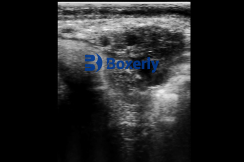
Ultrasound technology offers a real-time, non-invasive view inside the testicular tissue, providing both external size measurements and internal parenchymal evaluation. Typically, Modul B (brightness mode) ultrasound is used for this purpose. A linear transducer, usually in the 5–10 MHz range, provides high-resolution images of the testes.
Key Parameters Measured by Ultrasound:
1. Testicular Volume and Size
Testicular volume is one of the strongest predictors of sperm production. Using ultrasound, the length, width, and height of each testis are measured. The formula for testicular volume (length × width × height × 0.523) allows for accurate estimation of testicular mass.
2. Parenchymal Echotexture
The internal echotexture of testicular tissue should appear homogenous and fine-grained under normal conditions. Heterogeneous patterns or hypoechoic (dark) areas may indicate fibrosis, degeneration, mineralization, or other pathological changes that compromise sperm production.
3. Scrotal Circumference Correlation
While scrotal circumference measurement remains a standard field method, ultrasound offers a more detailed assessment by evaluating internal structures that scrotal tape cannot measure.
4. Vascular Flow (Advanced Doppler Ultrasound)
În unele cazuri, Doppler ultrasound is used to assess blood flow within the testicular artery. Adequate blood supply is essential for maintaining optimal testicular temperature and function.
Predicting Semen Quality Based on Ultrasound Findings
Numerous scientific studies have established a strong correlation between testicular size (particularly volume) and semen characteristics such as:
-
Sperm concentration
-
Total sperm output
-
Percentage of morphologically normal sperm
-
Sperm motility
Bulls with larger, structurally normal testes are statistically more likely to produce higher volumes of viable, motile sperm. Conversely, bulls showing irregular echotexture, calcifications, or hypoechoic lesions may suffer from subfertility or infertility.
In practice, ultrasound assessment allows for early identification of bulls that may not meet breeding standards, even before semen collection and analysis begin. This is especially valuable in young breeding bulls being evaluated for sale or selection.
Case Example: Ultrasound Use in a Commercial Bull Breeding Program
On many modern breeding farms, ultrasound scanning has become a routine part of bull evaluations. De exemplu, a commercial Angus breeding operation may scan all yearling bulls at 12 months of age before they enter breeding soundness evaluation (BSE) protocols.
Using a portable veterinary ultrasound scanner, the technician records:
-
Right and left testicular dimensions
-
Total testicular volume
-
Parenchymal uniformity
Bulls with optimal ultrasound findings are moved forward to semen collection. Bulls with suspicious ultrasound findings may be rechecked, provided time for recovery if illness is suspected, or culled if testicular abnormalities are confirmed. This dual approach increases the efficiency of selecting highly fertile bulls for breeding programs.
Advantages of Ultrasound Over Traditional Evaluation Alone
1. Non-Invasive and Repeatable
Ultrasound scanning can be performed repeatedly without discomfort to the bull. This allows for longitudinal monitoring of testicular development from puberty through adulthood.
2. Early Detection of Subclinical Problems
While scrotal circumference may not reveal internal lesions or fibrosis, ultrasound can detect early-stage abnormalities before they manifest as reduced semen quality.
3. Objective Measurements
Unlike manual palpation, ultrasound provides quantitative, visual data that can be stored, reviewed, and compared over time.
4. Useful for Young Bulls
For young bulls that have not yet started producing semen, ultrasound gives valuable predictive information about future sperm production potential based on testicular development.
5. Safe and Portable
Portable ultrasound machines like the BXL-V50 model make it easy to perform field evaluations directly on the farm, reducing stress and logistical challenges associated with moving animals to specialized facilities.
Limitations and Considerations
While ultrasound assessment is a valuable tool, it should not entirely replace traditional semen analysis. Some conditions may not manifest as structural testicular changes detectable by ultrasound. Factors like sperm motility, DNA integrity, and acrosome function still require laboratory evaluation.
Moreover, operator experience plays a significant role in obtaining accurate and consistent ultrasound measurements. Proper training in probe positioning, image interpretation, and equipment maintenance is essential for reliable data.
Integration with Overall Breeding Soundness Evaluation (BSE)
In many countries, BSE protocols are widely used to assess the fertility potential of breeding bulls. Ultrasound scanning fits seamlessly into these evaluations by complementing:
-
Physical examination (locomotion, vision, general health)
-
Scrotal circumference measurement
-
Semen collection and analysis
By integrating ultrasound findings into BSE, veterinarians and breeders gain a comprehensive, multi-faceted view of bull fertility.
International Adoption and Growing Popularity
Globally, the adoption of ultrasound technology in bull fertility management is growing rapidly. Studies from North America, Europe, Australia, and South America increasingly document its value.
De exemplu:
-
In the United States, researchers at Kansas State University’s Beef Cattle Institute have incorporated testicular ultrasound into their advanced BSE programs for seedstock operations.
-
In Australia, large-scale beef breeding programs use ultrasound for yearling bull selection, particularly in breeds like Brahman and Santa Gertrudis.
-
In Brazil, ultrasound is used extensively for both dairy and beef bull evaluations, given the tropical climate’s influence on heat stress and testicular function.
International breeding associations now recognize ultrasound data as valid supplementary information when making genetic selections for fertility traits.
Practical Tips for Ultrasound Assessment of Bull Testicles
-
Timing: Perform ultrasound evaluations during cooler parts of the day to minimize thermal effects on testicular measurements.
-
Preparation: Shave or wet the scrotal skin for optimal probe contact and image quality.
-
Consistency: Always scan both testes to identify asymmetry or unilateral defects.
-
Equipment: Use high-frequency linear transducers (7.5–10 MHz) for best resolution of testicular parenchyma.
-
Documentation: Record images and measurements for future reference and comparison.
Concluzie
Ultrasound scanning of breeding bulls offers a powerful, non-invasive, and highly informative method to assess testicular size and predict semen quality. When combined with traditional breeding soundness evaluations, it enhances reproductive management by allowing earlier selection decisions, reducing economic losses, and improving overall herd fertility.
As technology continues to advance and portable, user-friendly devices like the BXL-V50 become more accessible, more producers worldwide are integrating ultrasound into routine herd management. The ability to visualize testicular health in real time empowers farmers and veterinarians to make proactive, data-driven decisions that optimize reproductive efficiency, animal welfare, and profitability.
Reference Sources:
-
Brito, L. F. C., et al. (2012). “Use of ultrasonography for the assessment of reproductive function in male ruminants.” Reproduction in Domestic Animals, 47(Suppl 3), 34-39. https://doi.org/10.1111/j.1439-0531.2011.01951.x
-
Barth, Un. D., & Waldner, C. L. (2002). “Factors affecting breeding soundness classification of beef bulls examined at the Western College of Veterinary Medicine.” Canadian Veterinary Journal, 43(4), 274-284. https://www.ncbi.nlm.nih.gov/pmc/articles/PMC339423/
-
Kansas State University Beef Cattle Institute. (2023). “Bull Fertility Evaluation Using Ultrasound.” https://www.beefcattleinstitute.org/bull-ultrasound