Repeat breeder cows present a significant challenge to cattle producers worldwide. These cows, despite multiple inseminations, fail to conceive, causing economic loss due to extended calving intervals and reduced productivity. Among the many reproductive disorders implicated in repeat breeding, subclinical endometritis—a silent uterine inflammation without obvious clinical signs—has gained particular attention from veterinary researchers and farmers alike.

Detecting subclinical endometritis early and accurately is critical to improving reproductive efficiency. Veterinary ultrasound technology has emerged as a powerful, non-invasive tool that enables practitioners to diagnose this hidden condition in repeat breeder cows effectively. This article explores the role of veterinary ultrasound in detecting subclinical endometritis, its diagnostic advantages, practical applications, and how foreign research and farming practices have shaped current understanding.
Understanding Repeat Breeder Syndrome and Subclinical Endometritis
Repeat breeder syndrome (RBS) is commonly defined as the failure of a cow to conceive after three or more successive artificial inseminations (AI) or natural matings without obvious reproductive pathology. This syndrome affects approximately 10–15% of dairy and beef herds globally, leading to significant economic losses due to wasted semen, labor, and extended non-productive days.
Subclinical endometritis (SCE) refers to an inflammation of the uterine lining that lacks overt clinical signs such as discharge or fever. Unlike clinical endometritis, which can be visually diagnosed by vaginal discharge scoring, SCE is more elusive. It impairs the uterine environment, disrupting embryo implantation and reducing conception rates.
Early detection of SCE in repeat breeder cows is therefore essential to improve fertility outcomes. However, traditional diagnostic methods such as uterine cytology and biopsy are invasive, time-consuming, and not always practical on farms.
Veterinary Ultrasound Technology: A Game Changer in Detection
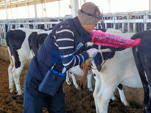
Veterinary ultrasound offers a non-invasive, real-time imaging method to examine the reproductive tract of cows. Transrectal ultrasonography, specifically, has become a standard technique in bovine reproductive management worldwide.
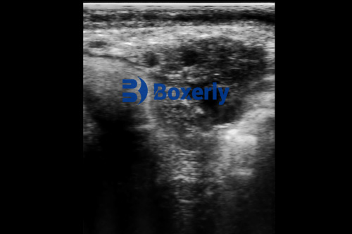
Using a high-frequency ultrasound probe inserted into the rectum, veterinarians can visualize the uterus, endometrium, and ovaries. The ultrasonographic appearance of subclinical endometritis includes:
-
Increased endometrial thickness
-
Presence of echogenic fluid or debris in the uterine lumen
-
Altered uterine wall echotexture reflecting inflammation
This technology allows for rapid screening of repeat breeder cows without stress or harm, facilitating timely interventions.
How Ultrasound Detects Subclinical Endometritis
Endometrial Thickness Measurement
One of the key ultrasonographic indicators of SCE is an increased thickness of the endometrium beyond the normal range. Studies from Europe and North America suggest that endometrial thickness exceeding 5 mm in the mid-diestrus phase correlates with uterine inflammation.
Veterinarians measure this thickness by placing calipers on the ultrasound screen at the inner and outer borders of the uterine lining. Repeat breeder cows with endometrial thickness above this threshold are flagged for further evaluation or treatment.
Detection of Uterine Fluid
Ultrasound can detect the presence of intrauterine fluid, which appears as hypoechoic (dark) or anechoic areas within the uterine lumen. While small amounts of fluid may be normal post-estrus, persistent fluid accumulation is a hallmark of endometritis.
Foreign research has emphasized the importance of fluid echogenicity: echogenic or cloudy fluid often indicates inflammatory debris or pus, which is consistent with subclinical infection.
Uterine Wall Echotexture Changes
Inflammation leads to subtle changes in the uterine wall’s echogenicity and texture. Instead of a smooth, homogenous endometrium, cows with SCE often exhibit heterogeneous patterns, with irregular or thickened areas visible on ultrasound scans.
Comparative Diagnostic Methods and the Role of Ultrasound
Traditionally, diagnosis of subclinical endometritis relied on cytological examination of uterine samples obtained via cytobrush or low-volume lavage. These techniques, while accurate, are invasive and require laboratory facilities for cell counting.
In contrast, veterinary ultrasound offers several advantages:
-
Non-invasive and animal-friendly: No physical sampling of uterine tissue is needed.
-
Immediate results: Ultrasound images are available in real time.
-
Repeatability: Ultrasound can be performed multiple times during the estrous cycle to monitor uterine health.
-
Cost-effectiveness: Avoids laboratory fees and reduces labor.
Studies from universities in Europe (e.g., University of Edinburgh, Wageningen University) and the United States (e.g., University of Wisconsin-Madison) have demonstrated good correlation between ultrasound findings and cytology results, validating ultrasound as a reliable screening tool.
Practical Applications on the Farm
Screening Repeat Breeder Cows
Farmers and veterinarians use ultrasound routinely to examine cows after failed inseminations. Detecting increased endometrial thickness or fluid accumulation can identify cows with SCE that may benefit from therapeutic interventions such as intrauterine antibiotics or hormonal treatments.
Timing of Treatment and Rebreeding
Ultrasound allows precise monitoring of uterine recovery after treatment. Cows with persistent signs of inflammation can be retreated or culled early, preventing prolonged infertility.
Integration with Fertility Programs
In modern dairy and beef operations, ultrasound diagnostics form part of comprehensive reproductive management programs. Alongside heat detection and ovulation synchronization protocols, ultrasound helps optimize breeding success rates and reduce days open.
Veterinary Ultrasound Innovations Enhancing SCE Detection
Recent advances in ultrasound technology have further improved the detection of subclinical endometritis:
-
Color Doppler Ultrasound: This modality evaluates blood flow in the uterine arteries. Increased vascularization may indicate inflammation. Studies show that color Doppler combined with B-mode imaging enhances diagnostic accuracy.
-
3D and 4D Ultrasound: While not yet widespread in large animal practice, 3D imaging offers volumetric assessment of the uterus, potentially allowing better detection of subtle lesions.
-
Portable and Wireless Ultrasound Devices: Compact units have increased field use, enabling on-site evaluation by veterinarians and trained farm personnel, even in remote locations.
International Perspectives and Research Insights
Across continents, research into subclinical endometritis and repeat breeder syndrome consistently highlights ultrasound as a cornerstone of diagnosis.
-
In Europe, where dairy production is highly intensive, routine post-partum ultrasound screening has been implemented to detect uterine health issues early.
-
In North America, ultrasound combined with cytology and bacteriology forms the basis of advanced reproductive health protocols.
-
In developing countries, ultrasound’s non-invasive nature makes it an attractive tool where laboratory infrastructure is limited.
Moreover, multinational studies emphasize that ultrasonographic parameters must be interpreted alongside clinical history, reproductive stage, and other diagnostics for best results.
Challenges and Limitations
While ultrasound is a valuable diagnostic tool, some limitations exist:
-
Operator dependency: Accurate diagnosis requires skilled veterinarians trained in bovine ultrasonography.
-
Interpretation variability: Slight differences in endometrial thickness cutoffs may occur based on breed and parity.
-
Cost of equipment: High-quality ultrasound machines may be cost-prohibitive for small farms.
Despite these challenges, ongoing training programs and technological advancements are addressing these issues.
Conclusion
Veterinary ultrasound technology has revolutionized the detection of subclinical endometritis in repeat breeder cows by offering a non-invasive, rapid, and effective diagnostic approach. This technology bridges the gap between clinical examination and invasive sampling, empowering veterinarians and farmers with actionable insights to enhance reproductive efficiency.
By identifying uterine inflammation early, treatment can be targeted precisely, reducing infertility, improving conception rates, and ultimately enhancing herd productivity and profitability. International research and field practices affirm ultrasound’s essential role in modern bovine reproductive management, promising a future where repeat breeding losses can be substantially minimized.
References and Sources
-
Sheldon, I.M., et al. (2009). Subclinical endometritis in dairy cattle: Pathogenesis and diagnosis.” Animal Reproduction Science, 121(3-4), 255-261. https://doi.org/10.1016/j.anireprosci.2010.04.013
-
Dubuc, J., et al. (2010). Evaluation of endometrial thickness by ultrasonography to diagnose endometritis in dairy cows.” Theriogenology, 74(9), 1509-1516. https://doi.org/10.1016/j.theriogenology.2010.06.028
-
Kasimanickam, R.K., et al. (2005). Endometrial cytology and ultrasonography for diagnosis of subclinical endometritis in dairy cows.” Theriogenology, 63(1), 818-830. https://doi.org/10.1016/j.theriogenology.2004.04.004
-
University of Wisconsin-Madison, School of Veterinary Medicine. (2023). “Use of Ultrasound in Bovine Reproductive Health.” https://www.vetmed.wisc.edu/repro
-
University of Edinburgh, Royal (Dick) School of Veterinary Studies. (2022). “Ultrasound diagnostics in dairy herd fertility management.” https://www.ed.ac.uk/vet/facilities/ultrasound