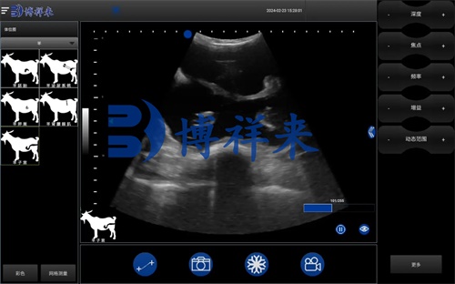
For goat farmers, accurate pregnancy diagnosis is critical for optimizing herd productivity, improving animal welfare, and ensuring efficient resource allocation. One particular area where early and accurate diagnosis has tremendous value is in identifying twin pregnancies. Goats are known for their relatively high natural twinning rates compared to many other livestock species, but managing twins requires different nutritional, housing, and veterinary care protocols than single pregnancies. Among the available diagnostic technologies, ultrasound—specifically B-mode ultrasonography—stands out as a safe, reliable, and non-invasive tool for detecting twin pregnancies in goats. In this article, we will explore how ultrasound tools are used to diagnose twin pregnancies, why they are increasingly adopted on goat farms worldwide, and how they contribute to better herd management.

Understanding Twin Pregnancies in Goats
Goats, especially dairy and meat breeds such as Boer, Nubian, and Saanen, frequently deliver twins or even triplets. While multiple births can increase productivity and profitability, they also introduce risks such as:
-
Nutritional demands: Pregnant does carrying twins require more energy and protein, particularly during the second and third trimesters.
-
Birthing complications: The risk of dystocia (difficult labor) increases with multiple fetuses.
-
Postpartum care: Twin kids may have lower birth weights and require more intensive neonatal management.
Therefore, knowing early whether a goat is carrying twins allows farmers to adjust management strategies well in advance, reducing losses and improving survival rates.
Traditional vs. Ultrasound-Based Pregnancy Diagnosis
Before the widespread adoption of ultrasound, goat pregnancy diagnosis often relied on:
-
Behavioral observation: Monitoring for return to estrus after breeding.
-
Palpation: Manually feeling for uterine changes.
-
Hormonal assays: Measuring progesterone levels in blood or milk.
While these methods have some utility, they lack the precision and early-detection capability necessary for identifying twin pregnancies. For example, hormonal tests can confirm pregnancy but cannot distinguish between single and multiple fetuses. Palpation is subjective and often unreliable for multiple fetus counts, especially in early gestation.
Ultrasound tools, on the other hand, have revolutionized pregnancy diagnosis by providing real-time visualization of the uterus, embryos, and placental structures.

The Advantages of B-Mode Ultrasound in Goat Twin Diagnosis
B-mode (brightness mode) ultrasound generates two-dimensional cross-sectional images, allowing veterinarians and trained technicians to observe fetal structures inside the uterus directly. Some of the key advantages include:
-
Early detection: Twins can often be reliably detected as early as 30 to 40 days into gestation.
-
Accurate fetal counting: Ultrasound allows direct visualization and counting of embryonic sacs and developing fetuses.
-
Non-invasive and safe: The procedure causes no discomfort or harm to the doe or fetuses.
-
Real-time data: Instant feedback enables immediate management decisions.
On many farms in the United States, Australia, Europe, and New Zealand, portable ultrasound machines have become a standard part of reproductive management protocols for goats.
How Ultrasound Scanning is Performed
Ultrasound scanning for goat pregnancy diagnosis can be performed using two main approaches:
1. Transabdominal Scanning
-
Timing: Typically performed after 30 days of gestation.
-
Procedure: The transducer is placed on the shaved or alcohol-wetted lower abdomen of the doe.
-
Advantages: Easy to perform in standing animals; minimal restraint required.
2. Transrectal Scanning
-
Timing: Can detect pregnancies as early as 18-20 days post-breeding.
-
Procedure: The probe is inserted into the rectum to get close proximity to the uterus.
-
Advantages: Allows for earlier pregnancy detection but requires more skill and equipment.
In most commercial goat farms, transabdominal B-mode ultrasound is the preferred method for detecting twin pregnancies due to its simplicity and reliability after 30 days.
Interpreting Ultrasound Images for Twin Diagnosis
With proper training, technicians can easily distinguish between single and twin pregnancies. Key indicators include:
-
Number of embryonic sacs: Each fetus is typically encased in its own fluid-filled sac.
-
Presence of two or more fetuses: Direct visualization of multiple fetal heartbeats confirms twins.
-
Placental structure: Observing the separation of the fetal membranes can aid in accurate counting.
However, the skill of the operator is crucial. Mistaking a single fetus that moves between uterine horns for two separate fetuses is a common error among inexperienced users. This is why training and practice are emphasized in veterinary ultrasound education worldwide.

Case Studies from International Goat Farms
United States: Commercial Dairy Goats
In large dairy goat operations in California and Wisconsin, ultrasound is routinely used not only to confirm pregnancies but also to count fetuses. By identifying twin pregnancies early, these farms adjust feed rations during late gestation to prevent pregnancy toxemia, a potentially fatal condition common in multiple-bearing does.
Australia: Meat Goat Enterprises
Australian Boer goat farmers frequently use portable ultrasound devices to identify twin pregnancies before the dry season. By separating twin-bearing does, they can prioritize access to higher-quality forage and supplements, ensuring strong birth weights and reducing kid mortality.
Europe: Hobby and Commercial Breeders
In countries like France, Spain, and the UK, small-scale hobby breeders as well as large commercial goat dairies incorporate B-mode ultrasound in routine herd management. European veterinary associations regularly offer training workshops to help goat farmers develop competence in ultrasound scanning.
Economic Benefits of Twin Pregnancy Detection
From an economic standpoint, early twin diagnosis provides multiple advantages:
-
Optimized nutrition: Feeding twin-bearing does according to their elevated nutritional needs improves fetal growth and maternal health.
-
Labor planning: Knowing the likely birthing difficulty allows farmers to schedule additional labor or veterinary assistance during kidding.
-
Reduced mortality: Proactive management reduces perinatal losses.
-
Efficient culling decisions: Does consistently producing singles may be culled or managed differently.
For commercial producers, these benefits directly translate into improved profitability and sustainability.
Limitations and Challenges
While ultrasound is a highly effective tool, some limitations exist:
-
Operator dependency: Accurate fetal counting requires training and experience.
-
Equipment cost: While prices have decreased, high-quality machines still represent a significant investment.
-
Early-stage challenges: Before 30 days, fetal structures may be too small for reliable counting via transabdominal scanning.
-
False negatives or positives: Occasionally, embryonic loss after scanning may mislead subsequent management if repeat scans are not performed.
To address these challenges, many producers opt for follow-up scans in mid-gestation to confirm early findings.
Technological Advancements in Portable Veterinary Ultrasound
In recent years, advancements in portable veterinary ultrasound equipment have made these tools more accessible and user-friendly. Features that have contributed to their growing popularity include:
-
Lightweight design: Easily transported to remote pastures or mobile veterinary clinics.
-
Long battery life: Essential for use in field conditions.
-
High-resolution imaging: Improved visualization of small fetal structures.
-
Durability: Waterproof and dustproof models withstand farm environments.
Global veterinary equipment manufacturers continue to innovate in this field, developing systems specifically tailored for small ruminants like goats.

Veterinary Training and Global Adoption Trends
Across veterinary schools and continuing education programs in North America, Europe, and Australasia, ultrasound competency is now a core part of reproductive management curricula. Many countries have national certification programs that credential technicians in small ruminant ultrasound scanning. The international collaboration between veterinary schools and agricultural extension services has accelerated global adoption, especially in developing regions where goats are vital to food security and rural economies.
The Future of Ultrasound in Goat Reproductive Management
Looking ahead, several exciting trends are emerging:
-
Artificial Intelligence (AI) integration: Early AI-driven ultrasound software can assist inexperienced operators in fetal counting and anomaly detection.
-
Telemedicine: Remote ultrasound consultations allow field technicians to transmit images to veterinary specialists for real-time interpretation.
-
Data integration: Ultrasound findings are increasingly linked to digital herd management software, enhancing long-term breeding decisions.
These innovations promise to further enhance the precision, accessibility, and utility of ultrasound in goat herds worldwide.
Conclusion
In the modern goat farming industry, early and accurate detection of twin pregnancies is no longer a luxury—it’s a necessity. Ultrasound tools, particularly B-mode scanners, offer farmers a non-invasive, real-time, and highly reliable method for managing reproductive performance. From small family farms to large commercial operations, the adoption of ultrasound technology has improved animal welfare, boosted kid survival rates, optimized resource allocation, and increased overall profitability.
While challenges remain, ongoing technological advancements and global knowledge-sharing are making ultrasound scanning an essential skill for goat producers everywhere. As the industry evolves, those who embrace this technology will be best positioned to achieve sustainable, efficient, and successful goat production.
References
-
Gabor, G., Taverne, M.A.M., & Szenci, O. (2008). Pregnancy diagnosis in goats: review of current methods and effectiveness of ultrasonography. Small Ruminant Research, 75(2-3), 130-135. https://doi.org/10.1016/j.smallrumres.2007.08.008
-
Knights, M., & Garcia, G.W. (2021). Reproductive Management of Small Ruminants Using Ultrasonography. Journal of Animal Science and Biotechnology, 12(1), 95. https://jasbsci.biomedcentral.com/articles/10.1186/s40104-021-00606-3
-
International Goat Association (2024). Best Practices for Reproductive Management in Goat Production. https://www.iga-goatworld.com/reproduction.html