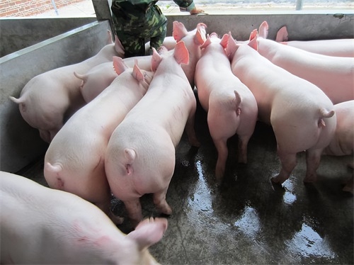
Ultrasound technology has become an indispensable tool in modern livestock production, especially in pig farming. It allows producers and veterinarians to assess internal structures non-invasively, providing vital information that can improve reproductive performance, animal welfare, and overall productivity. This article explores the main applications of ultrasound in pig farming, the benefits it brings, and the practical considerations for its effective use.
1. Basic Principles of Ultrasound in Pigs
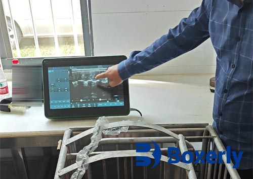
Ultrasound (or ultrasonography) uses high-frequency sound waves—usually between 2 to 10 MHz in livestock imaging—to visualize internal organs and structures. A transducer sends sound waves into the body, and the echoes reflected back are converted into images. These images are displayed in real time, allowing dynamic assessment of tissues.
The most common imaging mode used in pig farming is B-mode (brightness mode), which provides grayscale images of internal organs. For pregnancy detection and fat measurement, portable and waterproof machines are often used on farms, with probes designed for transabdominal scanning.
2. Reproductive Management
2.1 Pregnancy Detection
One of the most widespread uses of ultrasound in pig farming is early pregnancy detection. Ultrasound enables confirmation of pregnancy as early as 21–28 days after insemination. This early detection helps farmers quickly identify non-pregnant sows, allowing timely rebreeding and reducing days lost in production cycles.
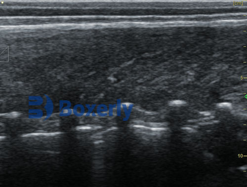
Real-time B-mode scanners show fluid-filled uterine structures, embryos, and fetal movements. As gestation progresses, more details—such as fetal skeletons—can be visualized, improving accuracy.
2.2 Evaluation of Reproductive Organs
Ultrasound is also used to assess the ovaries, uterus, and other reproductive structures. It can help detect abnormalities such as cystic ovaries, uterine infections, or delayed involution post-weaning. This enables veterinarians to implement targeted treatments and improves herd fertility management.
2.3 Determination of Litter Size
Although challenging, experienced operators may estimate litter size using ultrasound, especially after 28–35 days of gestation. While it is not always precise, such estimates help prepare facilities and optimize farrowing management.
3. Monitoring Body Condition and Backfat
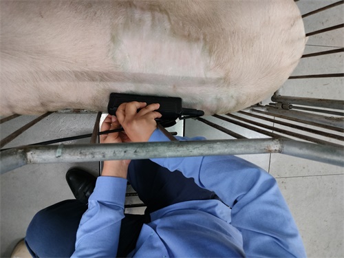
Proper body condition management in sows is essential for reproductive success and longevity. Ultrasound enables accurate measurement of backfat thickness—usually taken between the 10th and last rib, 6.5 cm off the midline.
This information is critical for feeding strategies during gestation and lactation. Overconditioned or underconditioned sows are more prone to farrowing difficulties, lower milk production, and decreased longevity. Using ultrasound ensures precise and individualized nutritional adjustments.
4. Carcass Quality and Genetic Selection
In commercial pig farming, ultrasound plays a key role in assessing carcass traits such as:
-
Backfat thickness
-
Longissimus dorsi (loin) muscle area
-
Intramuscular fat content
These traits are used for breeding stock selection and genetic improvement programs. Ultrasound provides live animal data, which is more ethical and cost-effective than post-mortem measurements.
Breeding companies and AI centers often use ultrasound data to predict meat quality and performance traits, which is valuable in developing high-yielding pig lines.
5. Health Monitoring and Disease Diagnosis
Ultrasound is also applied in veterinary diagnostics for pigs. Though less common than in companion animals, its use is growing in:
-
Detecting abscesses or internal fluid accumulation
-
Diagnosing umbilical hernias or scrotal swellings in piglets
-
Monitoring internal injuries in breeding boars or sows
Real-time imaging allows veterinarians to diagnose without invasive procedures, improving welfare and reducing antibiotic use.
6. Ultrasound-Guided Procedures
Advanced applications include ultrasound-guided biopsies or fluid aspirations. Though not routinely used in all farms, these techniques are becoming more accessible, especially in high-value breeding animals.
Ultrasound guidance increases accuracy and reduces the risk associated with blind punctures, making procedures safer for both animals and handlers.
7. Practical Considerations
To maximize the benefits of ultrasound, farmers must consider the following:
-
Training: Operators should be trained to interpret images accurately. Misdiagnosis can result from poor image quality or incorrect technique.
-
Equipment Maintenance: Probes and screens must be cleaned and maintained. Transmission gel is essential for image clarity.
-
Animal Handling: Calm and properly restrained animals yield better images. Stress can interfere with scanning and pose safety risks.
-
Biosecurity: Transducers should be disinfected between animals to avoid cross-contamination.
8. Future Developments
With advancements in imaging resolution, portability, and artificial intelligence (AI), ultrasound in pig farming is becoming more sophisticated. Some companies now offer AI-powered pregnancy detection or automated fat thickness measurement, reducing the need for manual interpretation.
3D imaging and Doppler ultrasonography, although not yet widespread in pigs, may become more common for advanced applications like vascular assessment or detailed reproductive evaluation.
Conclusion
Ultrasound is a highly valuable tool in modern pig farming, offering non-invasive insights into reproductive status, body condition, and health. Its use contributes to better animal management, increased productivity, and improved welfare. As technology continues to advance, ultrasound will likely play an even more integral role in sustainable swine production.
References
-
“Ultrasound in Swine Reproduction”, The Pig Site. https://www.thepigsite.com/articles/ultrasound-in-swine-reproduction
-
“Backfat and Muscle Depth Measurement Using Ultrasound”, Iowa State University Extension. https://www.extension.iastate.edu/news/2007/jan/071201.htm
-
“Using Ultrasound in Swine Management”, National Hog Farmer. https://www.nationalhogfarmer.com/health-wellness/article/21151771/using-ultrasound-in-swine-management