Hey everyone, I want to talk about how ultrasonography—or “ultrasound” for animals, like livestock and pets—gets used in monitoring growth, body condition, reproduction and respiratory health. I’ll share perspectives from overseas researchers and vets so you get a well‑rounded view. I’ve written this like a friendly chat, not a stiff research paper.
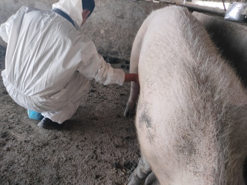
Imagine you’re working with cattle, sheep or goats and you wonder: how do we non‑invasively check how muscles are growing, how much fat they have, whether they’re pregnant, or if there’s a lung problem? Ultrasound is a tool many producers and vets abroad increasingly rely on. I’ll walk through key uses, how foreigners approach it, and why it’s become popular in many parts of the world.
Monitoring growth and carcass traits
In beef cattle especially, ultrasound has been around for decades to measure carcass traits in live animals. It helps predict things like rib fat, rump fat thickness, eye muscle area (EMA) and even intramuscular fat (marbling) before slaughter.
For example, Angus producers in Lithuania used ultrasonography on about 236 bulls and 22 heifers, linking weight gain with loin area, loin thickness and intramuscular fat. It turned out that once bulls hit about 431 kg and heifers about 603 kg, marbling was reliably detected—meaning ultrasound predicts meat quality quite well.
Other international studies compare ultrasound systems like Central Ultrasound Processing (CUP) versus Pie Medical Esaote Aquila (PIE) for predicting carcass traits. Findings show CUP often gives higher heritability estimates for intramuscular fat and better genetic correlations—so from a breeding perspective, the system choice matters.
Reproductive applications
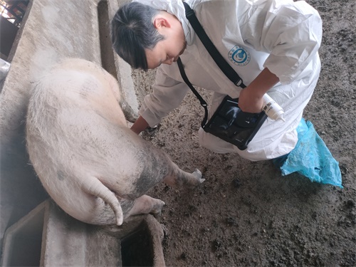
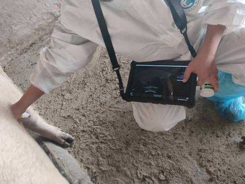
From a reproductive standpoint, real‑time B‑mode ultrasound and Doppler ultrasound have become essential tools. Foreign vets and researchers use ultrasound to diagnose pregnancy as early as 20–30 days, to monitor ovarian follicles, to determine fetal sex between day 55 và 85, and to assess embryo viability.
Color Doppler ultrasound goes one step further by showing blood flow in the corpus luteum or endometrium, which helps predict early pregnancy success and resynchronization approaches in beef and dairy herds.
Respiratory and thoracic health
On a different front, thoracic ultrasound (TUS) is gaining traction overseas for diagnosing bovine respiratory disease. Compared with traditional auscultation or visual signs, ultrasound reveals lung consolidations, pleural effusions, interstitial disease early—even before animals show clinical symptoms. Studies find it more accurate and prognostic, helping predict outcomes and guide interventions.
How people abroad actually use it—practical vibe
Foreign producers often talk about using ultrasound as part of routine herd checks. They’ll scan yearling steers or heifers at certain weights to guide finishing decisions, or ultrasound breeding candidates early in the breeding season to identify non‑pregnant or open cows quickly. This saves time, improves reproductive efficiency and boosts profits.
Veterinarians I’ve seen in international case studies emphasize that even novice operators can get consistent results, as long as they’re trained in probe placement and anatomy. That’s important—because poor training reduces diagnostic reliability.
Let’s talk key parameters—here’s a little table in HTML:
| Parameter | What it tells you | Typical use |
|---|---|---|
| Eye muscle area (EMA) | Muscle growth, meat yield | Genetic evaluation, finishing decisions |
| Subcutaneous fat thickness | Fat deposition, finishing readiness | Market timing, feeding plan |
| Intramuscular fat (IMF or marbling) | Meat quality, tenderness | Breeding & premium markets |
| Thoracic signs (lung consolidation, pleural fluid) | Respiratory disease presence/severity | Early diagnosis, treatment planning |
| Follicle or corpus luteum blood flow (Doppler) | Reproductive health, pregnancy potential | Breeding management, embryo transfer |
Growth patterns and the S‑curve vibe
Foreign animal scientists often describe livestock growth as an S‑shaped curve: after birth, growth is slow; then it accelerates until around puberty; eventually it plateaus as mature weight is reached and fat deposition dominates. Ultrasound allows producers to pinpoint where each animal is on that curve by measuring EMA and fat thickness over time.
When muscle growth slows and fat accumulation picks up, that’s usually the right time to switch feeding strategy—from lean‑mass production to finishing rations for marbling. Without ultrasound, farmers rely on guesswork, but abroad many have found that ultrasound-guided decision‑making improves uniformity and profitability.
Advantages from global perspective
-
Non‑invasive and animal‑friendly. Doesn’t stress animals, allows repeated monitoring without harm.
-
Quantitative and precise. Provides numeric values producers and breeders can trust and compare.
-
Real-time and actionable. You scan, see results, make decisions on the spot.
-
Supports genetic programs. Helps improve carcass traits faster by selecting the right candidates early.
-
Helps fight disease early. In respiratory or reproductive health, ultrasound spots problems sooner than palpation or visual signs.
Cultural perspectives and adoption trends
In North America and Europe, use of animal ultrasound is quite advanced—industry standards, genetic evaluation programs, veterinary training emphasize it. In Asia Pacific (including Japan, India, etc.), ultrasound adoption is growing rapidly too—especially in dairy and beef sectors, and even wildlife or endangered species research, where non‑invasive diagnostics matter.
Still, barriers remain: lack of equipment in some regions, limited operator training, and awareness gaps. Many foreign studies highlight that better training and awareness would boost adoption of ultrasound protocols in routine livestock practice.
Chatting about best practices
If you want to implement ultrasound in your own herd or clinic, here’s what foreign practitioners often recommend:
-
Invest in mid‑frequency real‑time B‑mode scanner (2–10 MHz) for carcass trait measures.
-
Train staff in anatomical landmarks: longissimus dorsi for EMA, intercostal spaces for rib/rump fat, thoracic ultrasound zones for lungs.
-
Use Doppler if reproductive flow assessment is needed—but it requires added skill and cost.
-
Scan at key stages: yearling entry to feedlot, mid‑finishing weight, pre‑breeding, early gestation (day 20–30) for reproduction.
-
Track and log measurements over time to build herd‑specific growth curves, differences by breed and feeding regimen.
Final thoughts
Across many countries, ultrasound of animals is more than a diagnostic novelty—it’s become a standard tool in precision livestock management. Whether you’re checking muscle growth, predicting meat quality, diagnosing pregnancy early, or catching lung disease before it spreads, it helps you make smarter decisions, saves money, and improves animal welfare.
I’ve leaned on real overseas studies and real experiences, so this isn’t just theory. From Lithuania to Australia, the U.S. to Japan, folks see the same thing: when ultrasound gets used thoughtfully, results follow.