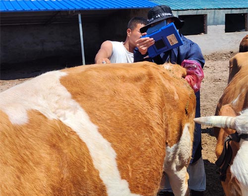
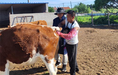
As a cattle farmer deeply involved in reproductive management, I have come to rely heavily on ultrasonography to monitor ovarian structures, particularly the corpus luteum (CL), during hormone-based interventions. One such intervention is the administration of prostaglandins (PGF2α), which play a crucial role in synchronizing estrus and improving breeding efficiency. Ultrasonographic imaging allows me to assess the structural and functional changes in the ovaries in real time, providing invaluable insights that directly inform my herd management decisions.
Trong bài viết này, I will share how bovine ultrasonography can be used to monitor the regression of the corpus luteum following prostaglandin treatment, drawing from both scientific principles and personal farming experience. The information here is especially useful for other livestock farmers seeking to improve reproductive outcomes through better understanding and timing of hormonal protocols.
Understanding the Role of the Corpus Luteum and Prostaglandins
The corpus luteum is a temporary endocrine structure that forms on the ovary after ovulation. Its main function is to produce progesterone, which is essential for maintaining pregnancy. If pregnancy does not occur, the body initiates a process called luteolysis—breakdown of the CL—primarily induced by prostaglandin F2 alpha (PGF2α), naturally produced by the uterus.
Veterinarians and farmers use synthetic prostaglandin injections to control the reproductive cycle, most commonly to synchronize estrus. However, the effectiveness of this treatment depends significantly on the timing within the estrous cycle when it is administered.
Timing Matters: When Does the CL Respond to Prostaglandins?
Prostaglandin treatment is only effective when the corpus luteum has matured sufficiently to be responsive. Through daily herd observations and routine ultrasonographic scans, I’ve noticed distinct patterns.
-
Days 5–6 After Estrus: Some cows begin to respond to PGF2α. The CL is still developing, and its regression may be partial or delayed.
-
Days 7–8 After Estrus: This is the most reliable window. The CL is fully functional and most cows show a clear response to treatment.
-
Day 17 of the Cycle: When cows are treated at this late stage, natural luteolysis may already be underway. The cow’s own prostaglandins initiate regression, and on ultrasound, the CL appears much smaller or nearly invisible.
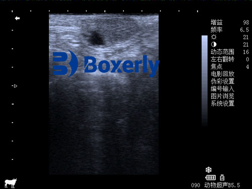
Using transrectal ultrasonography, I observe the CL shrinking in size and losing its typical echogenic texture as regression progresses. It becomes less distinct, often blending with surrounding ovarian tissue.
Follicular Waves and Their Impact on Ovulation Timing
The time from prostaglandin injection to ovulation isn’t fixed—it depends on the status of the ovarian follicles at the moment of treatment. Cows go through waves of follicle growth during the estrous cycle, with a dominant follicle selected in each wave.
-
Day 6 After Estrus (First Follicular Wave): If a dominant follicle from the first wave is still developing, prostaglandin treatment typically results in ovulation within 3 Ngày. I can visually track the follicle growing and maturing using B-mode ultrasound.
-
Day 13 After Estrus (Second Follicular Wave): At this stage, the second wave’s dominant follicle is just beginning to form. Ovulation tends to occur around 3.5 Ngày after treatment.
-
Day 9 After Estrus: Timing here is variable. In some cows, the first-wave follicle is still dominant and ovulates around Day 4 post-treatment. In others, the second-wave follicle is just initiating, and ovulation can be delayed to Day 6 post-treatment.
As a farmer, this variability is important to note. By integrating ultrasound exams with hormone protocols, I can predict ovulation windows with greater precision and improve the success rate of timed artificial insemination (TAI).
Visualizing Ovarian Changes via Bovine Ultrasound
B-mode (brightness mode) ultrasound is the standard imaging format I use on the farm. It creates cross-sectional grayscale images that allow me to view and measure ovarian structures.
When scanning the ovary:
-
A functional CL appears as a dense, rounded structure with a moderately echogenic (bright) texture.
-
After PGF2α treatment, I often see a gradual reduction in size. The CL begins to lose definition and becomes hypo-echoic (darker) as luteolysis sets in.
-
A mature follicle looks like a round, anechoic (đen) fluid-filled cavity, easily distinguishable from surrounding tissue.
Being able to differentiate between a regressing CL and an emerging follicle helps me make decisions about insemination timing and whether a second dose of hormone may be needed.
Effects of Cycle Stage on Conception Rates
My personal experience and veterinary literature both support the idea that the stage of the cycle at the time of prostaglandin treatment significantly affects fertility outcomes.
-
Early in the cycle (Day 5–6): Response is inconsistent. CLs may not regress fully, leading to failed ovulation or mistimed insemination.
-
Mid-cycle (Day 7–9): Optimal window for prostaglandin-induced luteolysis and synchronization. Conception rates are highest when insemination is timed accordingly.
-
Late-cycle (Day 17): Natural luteolysis may already be occurring. Using PGF2α here has limited impact, and fertility outcomes are mixed.
Kết thúc: Making the Most of Prostaglandin and Ultrasound Synergy
Ultrasonography has transformed how I manage reproduction in my herd. It bridges the gap between textbook theory and real-world variability in bovine physiology. By closely monitoring the corpus luteum and follicular dynamics after prostaglandin treatment, I can make informed decisions that improve breeding success, reduce open days, and ultimately increase profitability.
Understanding the intricate relationship between hormonal treatments and ovarian response is essential for anyone managing a cattle breeding operation. While prostaglandin protocols are powerful tools, they are most effective when guided by real-time ultrasonographic imaging and knowledge of the cow’s cycle stage.
References
-
Pierson, R.A. & Ginther, O.J. (1988). Ultrasonographic appearance of the bovine uterus during the estrous cycle. Sinh học.
-
Wiltbank, MC. et al. (2005). Prostaglandin F2α control of luteolysis and its timing in cattle. Animal Reproduction Science.
-
Fricke, Chiều. (2002). Ultrasonography for monitoring ovarian structures in dairy cows. Veterinary Clinics of North America: Food Animal Practice.