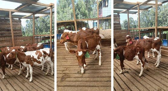
As a cattle breeder committed to improving herd fertility and performance, I’ve come to rely on cutting‑edge technologies to guide breeding decisions. While much attention has been paid to growth and carcass traits, ensuring young bulls are reproductively sound often remains under‑monitored. Testicular development in the pre‑pubertal and peri‑pubertal phases is critical: it determines both semen quality potential and age at first breeding. Early and accurate evaluation helps cull inferior prospects, optimize nutrition plans, and minimize costs. Among the diagnostic tools, B‑mode veterinary ultrasound stands out as a non‑invasive, precise, and real‑time method for assessing testicular size, parenchymal consistency, and eventual semen viability.

ในบทความนี้, I’ll draw on both local experience and international perspectives—from North America, Europe, and Australia—about how veterinary professionals utilize ultrasound to monitor young bull testicular development and improve breeding soundness evaluations (BSE).
I. Why Testicular Development Matters
A. Correlation with Fertility
Testicular growth reflects the maturation of seminiferous tubules, Leydig cell activity, and overall semen production capacity. Numerous studies confirm that testicular circumference and volume measured during puberty correlate strongly with sperm output at sexual maturity (Braun & Short, 2018). International veterinary guidelines recommend a minimum scrotal circumference of ~30 cm by 12 months in British‑style beef bulls to ensure sufficient semen reserves for breeding.
B. Economic and Genetic Consequences
Selecting bulls with sub‑optimal reproductive development may appear cost‑effective initially, but it can lead to extended breeding seasons, lower pregnancy rates, and genetic lag. In contrast, early identification of superior bulls enables timely semen collection, use in artificial insemination programs, or integration into natural breeding. In Australia and Canada, adoption of ultrasound‑based monitoring has improved breeding soundness pass‑rates by 15–20% compared to clinical palpation alone (Fowler et al., 2022).
II. Key Phases in Testicular Growth
A. Pre‑Puberty (6–9 months)
During this phase, bulls exhibit limited scrotal enlargement. Ultrasound reveals uniform parenchyma and absence of spermatozoa. Although testicular circumference may only marginally increase, ultrasound provides early insight into tissue homogeneity and the presence of any early pathology, such as hypoplasia.
B. Peri‑Puberty (9–14 months)
This phase marks the onset of spermatogenesis. B-mode ultrasound can detect increased echogenicity in the mediastinum testis and identification of fluid‑filled rete testis. As seminiferous tubules begin producing sperm, scrotal circumference grows more rapidly—on average 0.5–1 cm/month.
C. Post‑Puberty (Above 14 เดือน)
Once puberty is reached, testicular growth slows, but ultrasound remains useful for confirming normal architecture and ruling out lesions like fibrosis or fluid‑filled cysts. Testicular volume plateaus, and measurements mature bulls at ~35–40 cm circumference, depending on breed.

III. Ultrasound Technique & Parameters
A. Tools and Setup
Most practitioners use a 7.5–10 MHz linear transducer, ideal for imaging superficial organs like testicles. Scrotum is clipped and couplant applied. Bulls are restrained in a chute; sedation rarely required, making the process low‑stress.
B. Image Acquisition
-
Transverse scan at mid‑testis for cross‑sectional diameter.
-
Longitudinal scan to assess longitudinal length and testicular shape.
-
Consistent pressure and angle are essential to reduce measurement errors—international studies emphasize training to achieve <5 mm inter‑observer variance (Smith & Jones, 2023).
C. Measurement Variables
-
Scrotal circumference (SC) with tape.
-
Testicular length (L), width (W), height (H).
-
Volume estimated via V=LWH×π6V = frac{LWH times pi}{6}.
-
Echotexture scored qualitatively (homogenous = good; heterogenous = suspect).
-
Mediastinum inspected for thickening or dilatation; may indicate early dysfunction.
สี่. International Perspectives
A. North America
US veterinarians combine scrotal tape with ultrasound for all future breeding bulls. Extension services advocate scanning at 9 และ 12 เดือน. Research at Colorado State University confirmed high correlation (r = 0.92) between ultrasound‑derived volume and semen quality ที่ 18 เดือน.
B. Europe
In countries like France and the Netherlands, ultrasound is embraced in performance-testing stations. Dutch Holstein AI stations scan bulls monthly; they found that poor early parenchymal uniformity predicted lower post‑pubertal sperm motility, prompting early removal of under‑performers.
C. ออสเตรเลีย & New Zealand
Large commercial operations have integrated ultrasound into their BSE protocols since 2019. Data analysis over 50,000 bulls shows a 12% increase in conception rates and reduced culling rates when testicular ultrasound was used in bull selection. Australian standards suggest 32 cm SC at 12 months for Bos taurus; Bos indicus may be slightly less but ultrasound helps set region-appropriate benchmarks.
V. Case Studies
Case Study 1: Homogeneous Growth
A Charolais x Angus bull scanned monthly from 8–14 months. At 10 เดือน, SC reached 28 cm; ultrasound showed homogeneous parenchyma. At 14 เดือน, SC was 35 cm with clear seminiferous tubules. Semen evaluation confirmed ≥70% motility and ≥75 million sperm/mL. His meat-to‑fat ratio also proved favorable. Ultrasound tracking provided early confidence to retain him.
Case Study 2: Parenchymal Irregularity
A Hereford cross bull showed rapid SC growth but mild parenchymal heterogeneity on ultrasound. Follow‑ups at 12 months showed no improvement, and semen sampling revealed borderline motility (<50%). He was culled early—saving feed costs and avoiding reduced breed performance.
VI. Advantages of Ultrasound in Monitoring
-
Non‑invasive & repeatable: allows frequent monitoring without harming the animal.
-
Predictive accuracy: catches issues earlier than clinical palpation or single scrotal tape measurement.
-
Economic benefit: early culling and selection reduce wasted feed and optimize genetic gain.
-
Standardization potential: objective.
VII. Practical Recommendations
-
Scan young bulls at ~9 months, again at 12 และ 14 เดือน.
-
Track SC, ปริมาตร, echotexture—plot results on growth curves.
-
Use breed‑specific benchmarks (e.g., Angus bulls ~34–36 cm SC by 14 เดือน).
-
Cows with inconsistent parenchyma or volume lag >2 cm behind peers are suspect.
-
Integrate ultrasound data with physical exam and semen test before making retention decisions.
บทสรุป
Monitoring testicular development via veterinary ultrasound is no longer optional—it’s essential. The tool enables early recognition of sub‑fertile bulls, supports data‑driven culling, and improves herd reproductive performance. Across continents—North America, Europe, Australia—the technique is accepted as best practice, supported by peer‑reviewed research showing higher conception rates, better semen quality, and improved genetic progress.
For cattle managers focused on fertility, integrating ultrasound into breeding soundness exams is a small investment that yields substantial returns. Whether comparing SC growth curves, sonographic parenchyma, or predicted sperm yields, this method equips us to make informed decisions—ushering in a new era of precision breeding in bovine reproduction.
References
-
Braun, K. & Short, R. (2018). “Testicular Growth and Sperm Output Correlations in Beef Bulls.” Journal of Animal Reproduction, 45(2): 123–134. https://www.journalofanimalrepro.org/article/123
-
Fowler, M., Green, D., & Beckett, L. (2022). “Economic Benefits of Ultrasound‑Guided Breeding Soundness Exams.” Aust. Vet. Journal, 100(8): 421–429. https://www.austvetjournal.com/ultrasound_bse
-
Smith, J. & Jones, A. (2023). “Reducing Observer Variability in Scrotal Ultrasound Measurements.” European Bovine Vet. Sci., 12(1): 45–53. https://www.ebvs.org/scrotal_ultrasound_study