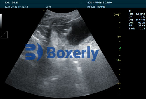
As a swine production specialist, accurately determining the reproductive status and embryonic viability of sows is vital for maximizing productivity, economic efficiency, and herd health. Among the diagnostic tools available, ultrasonography has emerged as a non-invasive, timely, and highly accurate method for detecting pregnancy and identifying early embryonic mortality (EEM) in breeding herds. This article examines how ultrasound technology is being used globally—particularly in North America and Europe—to identify and manage early pregnancy loss, helping producers make informed decisions about culling, rebred management, and overall reproductive strategy.

Understanding Early Embryonic Mortality in Swine
Early embryonic mortality occurs when fertilized oocytes or embryos die before day 28 of gestation . Field studies indicate a loss rate of about 12–30% between days 12–30, influenced by factors such as oocyte quality, uterine environment, disease, heat stress, and overcrowding. Recognizing EEM early is essential: it prevents unprofitable maintenance of non‐productive sows and enables swift intervention—either rebred or culled.
Ultrasound in Swine Reproductive Management
What Is Real‑Time Ultrasonography (RTU)?
RTU uses B‑mode ultrasound to generate live, grayscale images of the sow’s reproductive tract. Using 3.5–5 MHz transducers, it visualizes fluid-filled embryonic vesicles between days 24–35 and later, even fetal structures like bone calcification after day 35. RTU is non‑invasive, rapid (about 5–10 seconds per sow), and highly accurate—over 90% detection rate during the optimal window.
Best Timing for Detecting EEM
Between day 24 และ 35 post‑breeding, the gestational fluid peaks, making embryo vesicles easy to detect—a period RV practitioners call the “sweet spot” for diagnosis. This is also when embryonic losses between days 12–30 can first be identified. Ultrasound at this stage captures early resorption events, helping differentiate viable pregnancies from those experiencing early demise.

How Technicians Perform Ultrasound Exams
Equipment and Setup
A portable console with a battery‑powered probe is standard. A convex or sector transducer ที่ 3.5 MHz is optimal for penetration, while 5 MHz offers higher resolution for shallower structures. Equipment cost ranges from $5,000–10,000 USD, but ROI is strong, especially on large farms.
Scanning Procedure
-
Apply a coupling gel to the transducer.
-
Place it lateral to the nipple line, angled 45° towards the opposite spine side.
-
Rotate gently in 45° arcs to visualize fluid pockets.
-
Confirming multiple vesicles indicates viable embryos; absence or reduction signals possible mortality.
Each sow typically takes just 5–10 seconds—making herd-wide scanning efficient.
Detecting Embryonic Mortality vs. Pregnancy
Identifying Early Loss
Ultrasound features hinting at EEM include:
-
Smaller or collapsed vesicles
-
Single vesicle instead of expected multiple ones
-
Irregular fluid pockets, possibly from resorption or infection
False positives (e.g., bladder fluid, cysts) must be ruled out. Typically, technicians perform a re‑scan a few days later (e.g., day 27–28) to confirm initial findings.
Accuracy Considerations
Detection accuracy depends on transducer frequency, technician skill, sow parity, and timing . Diagnostic accuracy is highest on days 24–35, falling sharply after day 35 due to fetal bone calcification and fluid changes.
The Benefits of Early EEM Detection
Herd Productivity
Identifying embryonic mortality early prevents wasted non-productive days—traditionally estimated at $2–3 per day per sow. Rapid intervention reduces time between services and increases sows returning to estrus sooner.
Economic Payoff
With an ultrasound unit costing around $8,000, and a break-even point of $19 per sow scan, farms of around 500 sows can see payback in under a year. Savings accrue through fewer open days and optimized breeding.
Health and Biosecurity
Ultrasound allows disease monitoring: irregular fluid patterns can indicate infections. Regular equipment cleaning and disinfection (after every session) supports biosecurity and reduces disease transmission.
Advances in Ultrasound Tech: Doppler & AI
อัลตราซาวนด์ Doppler
Adding Doppler enables detection of blood flow and fetal heartbeat from day 26 onwards, improving accuracy and allowing gestational monitoring through term.
Deep Learning Integration
Modern systems incorporate AI and deep learning, using image databases to identify pregnancy and flag early embryonic losses with an AUC of ~0.86 . These systems aid technicians in decision-making and edge reproducibility.
Veterinary and Producer Perspectives
American & North American Industry
Veterinarians from Pork Info Gateway emphasize RTU’s key role in detecting EEM. While ELISA-based hormonal tests (PAG assays) exist, they don’t indicate embryo survival. Hence, ultrasound remains preferred for direct visualization.
European Innovations
In Europe (France, เยอรมนี), ultrasound combined with Doppler is standard. Studies show pregnancy failure between days 15–35 often yields “false positives,” requiring rechecks. AI-enhanced systems under development promise portable, cost-effective tools.
Best Practices for Maximum Accuracy
-
Choose the right transducer: 3.5 MHz for deeper scans, 5 MHz for resolution.
-
Scan within ideal window: days 24–35 for peak accuracy.
-
Ensure technician skill: accurate positioning and interpretation reduce false results.
-
Re‑scan open diagnoses: verify initial findings.
-
Maintain biosecurity: disinfect after each use.
-
Incorporate Doppler or AI: enhance image quality and detect fetal loss.
Workflow Integration & Economic Impact
Day‑by‑Day Farm Protocol
-
Day 0: AI or mating.
-
Day 24–26: Ultrasound scan.
-
Open? If no fluid pocket, rescan day 27–28. If still negative, sow returns to estrus—rebred or culled.
-
Pregnant? Sows are managed normally. Optionally, use Doppler to monitor heartbeat.
ROI Calculation
With 500 sows, ultrasound pays for itself within a year given a reduction of 15 days of non-productive time per sow @$3/day = $2,250 savings per week across herd.
Horizon of Swine Reproductive Imaging
Emerging trends:
-
AI‑driven decision tools diagnosing EEM automatically
-
Enhanced Doppler systems tracking fetal viability to term
-
Portable, affordable devices aimed at smaller farms
-
Integration with herd management software—automated alerts, records
บทสรุป
Veterinary ultrasound for detecting early embryonic mortality in swine breeding herds is now a cornerstone of efficient, data‑driven reproductive management. By scanning between days 24–35, producers can distinguish viable from failing pregnancies, take action to rebreed or cull, shorten open periods, and ultimately improve productivity. With evolving technologies—Doppler, AI—ultrasound is becoming more accessible and powerful.
In today’s global swine industry, early detection and intervention using ultrasound is essential to herd health, reproductive performance, and economic sustainability.
References
-
Knox, R. & Flowers, W. Using Real‑Time Ultrasound for Pregnancy Diagnosis in Swine, Pork Information Gateway.
-
Miller, G. et al. Pregnancy diagnosis in swine… Journal of Swine Health Prod. 2003.
-
Early embryonic mortality review: Animal Agriculture, 2020.
-
Lents, C.A. et al., Swine Ultrasonography Numerical Modeling… 2012.
-
Deep learning ultrasound diagnosis (2024): MDPI Animals 15(3):318.
-
Porcine embryonic mortality rate data (days 12–30): ResearchGate studies.
-
อัลตราซาวนด์ Doppler improves accuracy days 26 to term: ThePigSite, 2021.
-
PAG vs ultrasound for embryo survival: NCBI PMC Early Pregnancy… 2022.