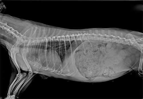
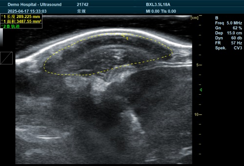
În medicină veterinară, diagnostic imaging plays a crucial role in identifying and managing various health conditions in animals. Two of the most commonly employed imaging modalities are radiography (X-rays) and ultrasonography (ultrasound). Each technique offers unique advantages and is suited for specific diagnostic purposes. This article delves into the fundamental differences between these two imaging methods, their applications, and considerations for their use in veterinary practice.

Radiography
What Is Radiography?
Radiography, commonly known as X-ray imaging, utilizes electromagnetic radiation to produce images of the internal structures of the body. When X-rays pass through the body, they are absorbed at varying degrees by different tissues, creating a contrast that is captured on a detector or film. Dense structures like bones absorb more X-rays and appear white, while softer tissues absorb less and appear in shades of gray. This imaging modality is particularly effective for evaluating:
-
Skeletal System: Detecting fractures, bone tumors, and joint abnormalities.
-
Thoracic Cavity: Assessing heart size, lung fields, and identifying conditions like pneumonia or heart failure.
-
Abdominal Cavity: Identifying foreign bodies, mărirea organelor, or gas patterns.
Radiography is a quick, non-invasive procedure that provides a broad overview of the body’s internal architecture. Însă, its ability to differentiate between soft tissues is limited.

Ultrasonography
What Is Ultrasonography?
Ultrasonography employs high-frequency sound waves to create real-time images of the body’s internal structures. A transducer emits sound waves that penetrate the body and reflect off tissues and organs. These echoes are then captured and transformed into visual images. Ultrasound is particularly useful for evaluating soft tissue structures, inclusiv:
-
Abdominal Organs: Liver, Rinichi, spleen, and intestines.
-
Cardiac Structures: Assessing heart function and detecting abnormalities.
-
Reproductive Organs: Monitoring pregnancies and diagnosing reproductive issues.
-
Guided Procedures: Assisting in needle biopsies or fluid aspirations.
Unlike radiography, ultrasound does not use ionizing radiation, making it a safer option for repeated evaluations. It provides detailed information about organ structure and function but has limitations in imaging areas containing gas or bone due to sound wave reflection.
Key Differences Between Radiography and Ultrasonography
| Aspect | Radiography (X-rays) | Ultrasonography (Ultrasound) |
|---|---|---|
| Imaging Principle | Uses electromagnetic radiation to capture images. | Utilizes high-frequency sound waves to produce images. |
| Best For | Evaluating bones, lungs, and detecting foreign objects. | Assessing soft tissues, organs, and guiding procedures. |
| Image Detail | Provides static images with good bone detail. | Offers real-time imaging with excellent soft tissue detail. |
| Radiation Exposure | Involves exposure to ionizing radiation. | No radiation exposure; safer for repeated use. |
| Limitations | Less effective for soft tissue differentiation. | Limited by gas and bone interference; operator-dependent. |
When to Use Each Modality
The choice between radiography and ultrasonography depends on the clinical scenario:
-
Radiography is preferred when assessing:
-
Bone fractures or joint issues.
-
Thoracic conditions like pneumonia or heart enlargement.
-
Detection of ingested foreign objects.
-
-
Ultrasonography is ideal for:
-
Evaluating abdominal organs for masses, Chisturi, or fluid accumulation.
-
Assessing heart function and detecting cardiac abnormalities.
-
Guiding fine-needle aspirations or biopsies.
-
În multe cazuri, both imaging modalities are used complementarily to provide a comprehensive diagnostic picture.

Concluzie
Radiography and ultrasonography are indispensable tools in veterinary diagnostics, each offering unique insights into an animal’s internal health. Understanding their differences, strengths, and limitations allows veterinarians to make informed decisions, ensuring accurate diagnoses and effective treatment plans. By leveraging the appropriate imaging modality, veterinary professionals can enhance patient care and outcomes.
References
-
“Radiography of Animals,” Merck Veterinary Manual.
-
“Ultrasound,” UC Davis School of Veterinary Medicine.
-
“Diagnosing Pets: Which Is Better, X-Ray or Ultrasound?” Castle Rock Animal Hospital.
-
“An Overview of Small Animal Veterinary Sonography,” Sage Journals.