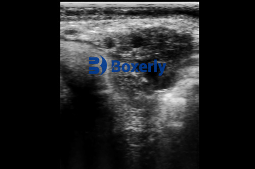
In modern livestock production, rapid, precis, and non-invasive diagnostics are essential for both economic success and animal welfare. Among the many tools available to veterinarians and cattle producers, ultrasound imaging has emerged as one of the most valuable. Particularly in dairy and beef herds, ultrasound allows for clear, real-time visualization of internal structures, supporting early diagnosis, effective reproductive management, and efficient herd health strategies. În acest articol, we explore how cattle ultrasound is used in clinical diagnosis and reproduction, its strengths and limitations, and why it’s gaining traction globally despite regional disparities in adoption.

Diagnostic Value of Ultrasound in Cattle
Early Detection of Internal Diseases
Ultrasound imaging provides a three-dimensional, real-time view of internal organs, making it possible to detect abnormalities before they become clinically visible. In cattle, this includes liver masses as small as 1 cm, abnormalities in the gallbladder, common bile duct, hepatic ducts, pancreas, adrenal glands, Rinichi, and prostate gland.
Compared to traditional palpation or biochemical tests, ultrasound allows the clinician to directly observe structural changes. De exemplu, in cases of hepatic abscesses or peritonitis, ultrasound not only helps detect lesions early but also aids in assessing their size, location, and contents. This precision helps veterinarians make faster and more effective treatment decisions.
Reproductive System Imaging
Ultrasound has transformed reproductive management in cattle. Through transrectal, transvaginal, or transabdominal approaches, veterinarians can assess ovarian structures, diagnose pregnancy at an early stage (as early as 28 Zile), and monitor fetal development. It is also instrumental in identifying reproductive disorders, including ovarian cysts, uterine infections, or structural deformities.
Ultrasound offers several advantages in obstetrics. It can help determine fetal position, detect twins, estimate amniotic fluid volume, locate the placenta, and identify developmental abnormalities like hydatidiform moles. These insights are critical for timely intervention, improving both calf survival and maternal health.

Reproductive Management Benefits
Enhancing Breeding Decisions
The ability to visualize follicles and corpora lutea with high clarity allows producers to synchronize estrus more effectively and increase conception rates through timed artificial insemination. This is especially valuable in intensive dairy systems where reproductive efficiency directly affects milk yield.
In countries like the United States, Canada, and New Zealand, ultrasound is a standard part of reproductive programs. Its application reduces the number of open (non-pregnant) days in dairy cows, saving substantial feed and labor costs.
Monitorizarea dezvoltării fetale
Fetal measurements such as crown-rump length, biparietal diameter, and heartbeat monitoring are achievable with real-time ultrasound. These measurements are essential not only for confirming viability but also for estimating gestational age and anticipating calving dates more precisely. In high-value pregnancies, such as embryo transfer programs, this is crucial for success.
Minimally Invasive and Repeatable
One of ultrasound’s most praised features is its non-invasive nature. It can be used repeatedly without harming the animal or affecting reproductive performance. This makes it ideal for herd-level monitoring in commercial settings.

Clinical Applications Beyond Reproduction
Detecting Fluid Accumulation and Cysts
Ultrasound excels at detecting fluid-filled structures. This includes ascites, cysts in the kidney or liver, or uterine fluid accumulation post-partum. The imaging not only reveals the presence of fluid but can also help characterize its physical nature—whether it’s clear, purulent, or hemorrhagic—informing more targeted treatments.
Calculating Volumes and Sizes
Veterinarians can measure the exact size and volume of abnormal structures. This is important when evaluating abscesses, hematomas, or tumors, and for monitoring their changes over time. Unlike palpation, ultrasound quantifies disease progression or regression, supporting evidence-based decision-making.
Urinary and Digestive System Imaging
Although air and bone limit the penetration of ultrasound waves, the bladder and kidney are accessible for evaluation. Conditions like nephritis, urolithiasis (bladder stones), or bladder rupture can be diagnosed early. In the digestive tract, ultrasound is useful for evaluating the reticulum in cases of traumatic reticuloperitonitis, even though full evaluation of gas-filled intestines remains challenging.
Limitations and Challenges
Limited Penetration Through Bone and Gas
One significant limitation of ultrasound is its poor ability to penetrate bone or air-filled structures. As a result, organs such as the lungs or gas-distended stomach are often difficult to evaluate. This makes ultrasound less useful than X-ray or CT for diagnosing pulmonary or skeletal diseases.
Tumor Detection Limitations
While ultrasound can detect many abnormalities, small tumors around 1 cm may still go unnoticed. A negative ultrasound result does not always exclude the presence of early-stage neoplasms. This is particularly important in cases involving subtle or early malignancies.
Artifacts and Misinterpretation
Ultrasound imaging relies on sound wave reflection, which can sometimes produce false images due to repeated echo patterns, side lobes, or shadowing. In inexperienced hands, these artifacts may lead to misdiagnosis. Therefore, training and experience remain critical components of accurate ultrasound interpretation.
Availability and Adoption
Despite its advantages, ultrasound use in cattle is not yet universal, particularly in parts of Asia, Africa, and Latin America. High equipment costs, limited training opportunities, and lack of awareness are major barriers. In China, for example, the adoption of cattle ultrasound began in academic institutions and select enterprises, but widespread use only gained traction in the last decade with the rise of commercial dairy operations and improvements in veterinary education.

Global Perspectives on Cattle Ultrasound
Veterinarians in countries like Germany, Australia, and the Netherlands have integrated ultrasound into standard herd health protocols. In the EU, government-backed animal welfare initiatives promote the use of non-invasive technologies like ultrasound to enhance productivity while minimizing stress and injury.
In the United States, the use of ultrasound in beef cattle has expanded beyond reproduction into carcass trait evaluation. Technologies like carcass ultrasound measure backfat thickness, eye muscle area, and marbling—parameters critical for genetic selection and meat quality grading.
In South America, especially in Brazil and Argentina, ultrasound is becoming a standard tool in embryo transfer and advanced breeding programs. Portable devices with robust performance and long battery life are favored due to the challenging environments and large herd sizes.
The Future of Ultrasound in Bovine Practice
Advances in ultrasound technology—such as Doppler imaging, 3D reconstruction, and portable wireless probes—are making the tool more versatile and user-friendly. Doppler technology, in particular, opens new diagnostic horizons by enabling real-time measurement of blood flow in small vessels, assessing tissue perfusion, and identifying vascular anomalies.
Integration with artificial intelligence and cloud-based data storage systems is also transforming herd-level diagnostics. AI-assisted interpretation can reduce diagnostic errors, improve consistency, and allow less-experienced users to generate reliable results.
With increasing global emphasis on sustainable livestock practices, ultrasound aligns with the goals of precision veterinary medicine. Its role in reducing antibiotic misuse, improving reproductive success, and minimizing economic loss due to late diagnoses makes it a cornerstone of 21st-century cattle health care.

Concluzie
Ultrasound imaging is revolutionizing how veterinarians and producers manage cattle health and reproduction. Its non-invasive nature, diagnostic accuracy, and growing affordability make it an indispensable tool in both intensive and extensive livestock systems.
While challenges such as penetration limitations and initial cost remain, the benefits—particularly in reproduction, internal medicine, and herd productivity—are undeniable. As education and access expand globally, ultrasound is set to become a routine part of veterinary practice in cattle industries around the world.
Reference Sources
-
Whitaker, D. A., & Smith, E. (2021). Veterinary Ultrasonography in Food-Producing Animals. Journal of Veterinary Imaging.
-
Beef Cattle Institute. (2023). “Use of Ultrasound in Cattle Diagnostics and Reproductive Management.” https://www.beefcattleinstitute.org/ultrasound-diagnostics
-
Lischer, C. J., et al. (2020). “Applications of Bovine Ultrasound in Reproduction and Internal Medicine.” Veterinary Clinics of North America: Food Animal Practice.