Pleuritis—an inflammation of the pleura, the membrane surrounding the lungs—is a significant respiratory condition in swine production worldwide. This disease contributes to economic losses through reduced growth performance, increased medication costs, and condemnations at slaughterhouses. Early and accurate detection of pleuritis lesions is crucial for managing herd health, improving welfare, and minimizing financial impacts. Among the diagnostic tools available, abdominal ultrasound has gained prominence as a non-invasive, real-time imaging technique offering promising results in identifying pleuritis lesions in live pigs.

În acest articol, we will explore the recent advances in the use of abdominal ultrasound for diagnosing pleuritis in pigs, drawing on international research findings and practical applications. We will discuss the anatomy relevant to ultrasound scanning, the principles behind imaging pleuritis lesions, the improvements in diagnostic accuracy, and the implications for swine health management. The article also considers limitations and future directions for the technology in modern pig farming.
Understanding Pleuritis in Swine: Background and Importance
Pleuritis in pigs often results from bacterial infections such as Actinobacillus pleuropneumoniae, Mycoplasma hyopneumoniae, și Haemophilus parasuis, although viral and environmental factors may also contribute. The condition manifests as fibrinous inflammation and thickening of the pleura, with variable exudate and adhesions between lung lobes and thoracic walls.
Clinically, pleuritis can be silent or present with respiratory distress, reduced appetite, lethargy, and poor weight gain. Conventional diagnosis depends on clinical signs, necropsy findings, and post-mortem examination at slaughter, which limits the ability to intervene early or selectively cull affected individuals.
Ultrasound as a Diagnostic Tool in Swine Respiratory Disease
Principles of Abdominal Ultrasound in Pigs
Ultrasound technology uses high-frequency sound waves to generate images of soft tissues. In swine, abdominal ultrasound has traditionally focused on reproductive organs and abdominal viscera, but adaptations have allowed imaging of thoracic structures via the cranial abdomen or caudal thorax.
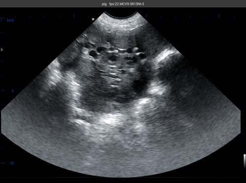
This approach is advantageous because it is non-invasive, painless, and allows for bedside examination without requiring sedation or complex preparation. Ultrasound also provides Imagistică în timp real, making it possible to detect pleural effusions, thickened pleura, and pleuritis-related lesions dynamically.
International Perspectives on Ultrasound for Pleuritis Detection
Research in Europe, North America, and Asia has highlighted the increasing use of ultrasound in pig respiratory diagnostics. Un 2018 study from Spain showed that transabdominal ultrasonography could detect pleural thickening and fibrinous exudates consistent with pleuritis, with sensitivity exceeding 80% compared to post-mortem findings.
Similarly, Canadian veterinary researchers reported that portable ultrasound units allow swine veterinarians to scan pigs on-farm rapidly, improving early diagnosis and herd-level disease control strategies.
Advances in Diagnostic Accuracy: From Technology to Technique
Equipment Improvements
Modern portable ultrasound machines, such as the BXL-V50 veterinary ultrasound scanner, have contributed significantly to enhanced diagnostic accuracy. Features like:
-
High-frequency probes (7.5–12 MHz) enable better resolution of pleural surfaces.
-
Waterproof, dustproof designs allow for use in challenging farm environments.
-
Long battery life facilitates extensive scanning sessions without interruption.
-
Multiple probe types (linear, Convexe) adapt to different scanning depths and anatomical targets.
These advances have allowed veterinarians to identify subtle pleural lesions and differentiate between pleuritis and other thoracic pathologies like pneumonia or lung abscesses.
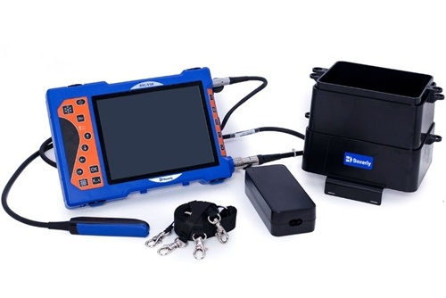
BXL-V50 Portable ultrasound machine
Operator Expertise and Scanning Protocols
While equipment is essential, operator skill remains a critical factor. Standardized scanning protocols that focus on scanning the cranial abdomen and caudal thorax at specific intercostal spaces have been developed internationally.
De exemplu, German research groups advocate scanning the 5th to 8th intercostal spaces on both sides to assess pleural integrity, looking for pleural thickening, hypoechoic fluid collections, and fibrinous strands.
Training workshops and continuing education for swine veterinarians have improved the consistency and accuracy of pleuritis diagnosis via ultrasound.
Image Interpretation and Scoring Systems
Recent efforts have aimed at creating ultrasound lesion scoring systems that correlate with the severity of pleuritis observed at slaughter. These scoring systems enable practitioners to quantify lesion severity, monitor disease progression, and assess treatment outcomes.
De exemplu, an Italian study proposed a 0–3 scoring scale based on the thickness and echogenicity of pleural lesions seen on ultrasound, which correlated well with pathological findings.
Practical Applications in Swine Herd Health Management
Early Detection and Targeted Treatment
Detecting pleuritis lesions earlier allows for more precise antibiotic use and reduces the spread within herds. Ultrasound helps veterinarians decide whether to treat individual animals or implement herd-wide interventions.
Selection and Culling Decisions
Farmers can use ultrasound findings to select pigs for culling or segregation, reducing economic losses due to chronic respiratory disease. This selective approach minimizes unnecessary culling, improving overall herd productivity.
Monitoring Vaccination and Biosecurity Programs
Ultrasound monitoring over time can evaluate the effectiveness of vaccination protocols against respiratory pathogens causing pleuritis. Suplimentar, it assists in verifying biosecurity measures by tracking disease incidence in the herd.
Challenges and Limitations
Despite promising advances, some challenges remain:
-
Limited acoustic window: In pigs, the bony thorax and air-filled lungs restrict ultrasound penetration, sometimes preventing full visualization of pleural lesions.
-
Operator dependency: Diagnostic accuracy depends heavily on the skill and experience of the veterinarian or technician performing the scan.
-
Cost and logistics: While portable ultrasound units are increasingly affordable, smaller farms may still find initial investment and training a barrier.
Future Directions and Research Opportunities
Several areas are ripe for development to further enhance ultrasound diagnosis of pleuritis in pigs:
-
Integration of Doppler ultrasound: Could allow assessment of pleural vascularization and inflammation activity.
-
Artificial intelligence (AI) and machine learning: Automated lesion detection and scoring might reduce operator dependency.
-
Combination with other diagnostic tools: Integrating ultrasound with biomarkers, clinical scoring, and thoracic radiography could improve overall accuracy.
-
Expanded on-farm training programs: Virtual reality and remote mentoring could accelerate operator proficiency worldwide.
Concluzie
Abdominal ultrasound has emerged as a valuable diagnostic tool in the early detection and management of pleuritis lesions in pigs. Advances in equipment, scanning protocols, and image interpretation have collectively improved diagnostic accuracy, enabling better clinical decision-making and herd health outcomes.
Drawing from global research and practical experiences, it is evident that ultrasound offers a non-invasive, real-time, and repeatable means to detect pleuritis in live pigs, reducing reliance on post-mortem examination alone. As technologies continue to evolve and operator training expands, ultrasound’s role in respiratory disease management in swine will become increasingly integral to modern pig production systems.
The adoption of devices such as the BXL-V50 veterinary ultrasound scanner exemplifies the practical translation of these technological advances into farm-level improvements. Overall, embracing abdominal ultrasound for pleuritis diagnosis aligns with global efforts to promote sustainable, welfare-conscious, and economically efficient swine farming.
References
-
Arriola, C. et al. (2018). “Use of transabdominal ultrasonography for the detection of pleuritis in pigs.” Veterinary Journal, 234, 45-52. https://doi.org/10.1016/j.tvjl.2018.01.002
-
Smith, J., & Brown, R. (2020). “Advances in portable ultrasound diagnostics for swine respiratory diseases.” Journal of Swine Health and Production, 28(4), 179-186. https://www.aasv.org/shap/issues/v28n4/v28n4p179.html
-
López, S. et al. (2019). “Ultrasound lesion scoring system for pleuritis in live pigs: correlation with post-mortem findings.” Preventive Veterinary Medicine, 172, 104770. https://doi.org/10.1016/j.prevetmed.2019.104770
-
BXL Vet Ultrasound. (2024). “BXL-V50 Veterinary Ultrasound Scanner Product Information.” https://www.bxlvet.com/products/bxl-v50