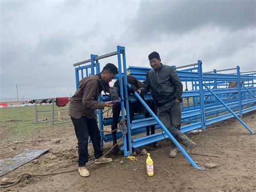
At horse breeding farms, spotting trouble early is the name of the game—especially when it comes to colic. Any equine vet will tell you, colic is a word no horse owner wants to hear. It’s not a disease but a symptom of abdominal discomfort, and the causes range from mild gas buildup to severe intestinal twists. For mares, especially those in late gestation or just post-foaling, colic can be life-threatening. But here’s the good news: portable ultrasound is changing the game, giving farm teams and vets a reliable, non-invasive way to track signs of colic before things spiral out of control.

Colic on Breeding Farms: Why It’s a Big Deal
On a breeding farm, every mare matters. Each one is a genetic investment, not to mention the potential value of the foal she’s carrying. That makes colic not just a health crisis—but an economic one too. Breeding mares are at higher risk due to late-pregnancy displacement of abdominal organs, post-foaling uterine changes, and diet-related shifts. Add the stress of environmental changes or herd dynamics, and you’ve got a perfect storm.
One of the biggest challenges with equine colic is that it often hides in plain sight. Early symptoms—like restlessness, decreased appetite, or a slight increase in heart rate—are easy to miss. That’s where ultrasound steps in. With a skilled operator and a good portable machine, subtle internal changes can be visualized and tracked, giving vital clues before the mare shows dramatic symptoms.
What Can Ultrasound Really See?
Let’s get one thing straight: ultrasound can’t diagnose every case of colic. But it can give vital information that shapes how a vet responds. A quick scan of the abdomen can reveal:
-
Intestinal distension from gas or fluid
-
Displacement or lack of intestinal motility
-
Uterine position in pregnant mares
-
Presence of abnormal peritoneal fluid
-
Impacted colonic segments
When scanning a mare showing vague signs—maybe she’s not finishing her feed or she seems uncomfortable after rolling—ultrasound offers a peek into what’s going on without needing sedation or invasive probes. Most importantly, it can help differentiate between mild colic that can be managed on-site and severe cases needing surgery.
Everyday Use: How Farms Are Putting Ultrasound to Work
Across the U.S., UK, and Australia, equine-focused farms are starting to include ultrasound in their routine checks, especially during foaling season. Some breeding farms have even trained barn staff to perform basic scans under veterinary guidance—enough to flag abnormalities for the vet’s attention.
One popular setup is using a portable B-mode ultrasound with a low-frequency convex probe, ideal for deeper abdominal imaging. Operators can assess motility in small intestines, visualize the colon’s gas pattern, and detect abnormal pockets of fluid. For late-term pregnant mares, scanning the uterine environment gives peace of mind and can catch early signs of uterine torsion or placental separation.
In Australia’s Hunter Valley, for instance, one large stud farm invested in a shared ultrasound unit for night watch teams. The result? Fewer emergency vet calls, earlier interventions, and improved outcomes in both mares and foals.
Not Just for Crisis: A Preventative Tool
Many horse owners think of ultrasound as something you use once a horse is already sick. But smart farms are flipping that mindset. They’re using ultrasound as a routine tool—especially for mares prone to colic or those with previous abdominal surgeries. A quick scan once a week takes just a few minutes and provides data points over time. If bowel loops start to enlarge or fluid patterns change, intervention can happen early, sometimes before the horse shows visible symptoms.
Ultrasound also offers peace of mind. เช่น, when a mare acts uncomfortable after foaling, it’s easy to panic. Is it retained placenta? A twisted uterus? Gas colic? With ultrasound, answers come faster—and treatments can be more targeted.
Ultrasound During the Peripartum Period
Late pregnancy and the days right after foaling are prime times for colic. The mare’s abdomen is undergoing major changes, and the GI tract often doesn’t keep up. Uterine torsion, uterine rupture, or uterine artery rupture can mimic colic and are difficult to differentiate without imaging.
Here, ultrasound can:
-
Confirm the fetus’s position in late gestation
-
Identify large uterine vessels or hematomas
-
Detect free fluid in the abdomen post-foaling
-
Assess bowel motility during postpartum recovery
Having an ultrasound available helps avoid unnecessary delays. While rectal palpation is still valuable, it’s not always enough—especially in smaller mares or early cases. And unlike rectal exams, ultrasound carries zero risk of rectal tear, making it a safer option in some scenarios.
Challenges and Limitations
Ultrasound isn’t magic. It’s operator-dependent, and image interpretation takes training. Abdominal gas can obscure images, and deep structures may not always be visible. Not every farm has access to skilled ultrasound operators or high-end machines. อย่างไรก็ตาม, even basic images can guide decisions when interpreted with clinical signs.
Some farms worry about cost. But portable machines are now more affordable than ever, and the long-term savings—fewer emergency calls, better outcomes, less downtime—more than make up for it. Plus, many practices now offer rental or shared-use equipment during breeding season.
What Vets Are Saying
Dr. Nicole Frazier, an equine vet based in Kentucky’s Bluegrass region, shared that “before portable ultrasound became part of our daily toolkit, we’d lose critical time in colic evaluations—especially in postpartum mares. Now we scan on the spot. We’ve caught early uterine artery bleeds, gas buildup, and even some odd bowel displacements without needing to haul to a clinic.”
European practices echo the sentiment. In Germany and the Netherlands, several equine clinics offer mobile ultrasound screening during foaling season. It’s part of a proactive health approach that fits well with modern, welfare-focused breeding.
The Bottom Line: Early Detection Saves Lives
When it comes to colic, time is everything. Horses are stoic animals—they often hide pain until it’s too late. Ultrasound bridges that gap, giving farm managers and vets a clearer view of what’s happening internally.
It’s not just about crisis management. When used proactively, ultrasound becomes a powerful part of a horse’s ongoing care—especially at breeding farms where reproductive health and abdominal stability go hand in hand.
If you’re working on a breeding farm and haven’t yet added ultrasound to your toolkit, it’s time to reconsider. Because when it comes to colic, seeing is believing—and sometimes, it’s life-saving too.