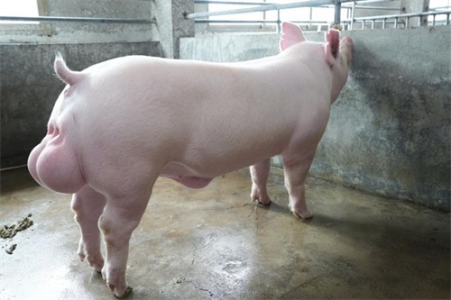
As a modern swine producer or animal scientist, gaining accurate insight into the body composition of breeding pigs—particularly intramuscular fat (IMF)—has become essential for genetic selection, nutrition optimization, and meat quality forecasting. Among the various available technologies, veterinary ultrasonography (commonly referred to as ultrasound) has emerged as a non-invasive, คุ้มค่า, and highly practical tool for evaluating intramuscular fat in live pigs.

Intramuscular fat, also known as marbling, plays a significant role in pork palatability, tenderness, juiciness, and overall eating experience. อย่างไรก็ตาม, measuring IMF in live pigs poses challenges due to its subtle deposition patterns and lower concentration compared to species like cattle. This article walks you through the essential preparatory steps and image quality standards when using อัลตราซาวนด์สัตวแพทย์ to assess IMF in breeding pigs, drawing on international best practices and practical experiences from farms and labs worldwide.
Why Intramuscular Fat Matters in Swine Production
IMF content is not merely a cosmetic trait—it is a fundamental determinant of pork flavor and market value. In many Western and Asian markets, marbled pork fetches higher prices, making it a breeding objective in both commercial and heritage swine genetics programs.
นอกจากนี้, IMF content is genetically influenced but also highly dependent on nutrition, age, and sex. Therefore, tracking it throughout the animal’s development using non-invasive methods like ultrasound allows producers to make data-driven decisions for selection and management.
Preparation Before Ultrasound Scanning
-
Restraining the Pig for Stability
The accuracy of any ultrasound scan begins with proper animal positioning. ในทางปฏิบัติ, pigs are gently restrained using metal crates or holding pens, allowing them to stand naturally without stress or struggle. International studies, including those from European swine research facilities, stress the importance of reducing movement, which can distort ultrasound images and affect reliability.
Unlike in cattle or small ruminants, pigs are often less tolerant of lengthy handling, so the process must be quick, efficient, and humane.
-
Identifying and Preparing the Scanning Area
Once the pig is restrained, the next step is identifying the proper scanning location. Based on global consensus, three anatomical regions are most commonly used:
-
The last rib and 3rd–4th lumbar vertebrae area
-
The 10th rib position
-
The last lumbar vertebra
Experienced technicians can locate these landmarks by palpation. Prior to scanning, the hair on the scanning site is clipped, and any remaining stubble is cleared to ensure optimal contact between the probe and skin. This is critical—studies from North American swine herds indicate that even minimal hair interference can compromise image clarity.
-
Application of Ultrasound Coupling Gel
ต่อไป, a generous amount of ultrasound gel is applied to the scanning site. The gel eliminates air gaps between the transducer and pig’s skin, which can otherwise obstruct the transmission of sound waves. In countries like Germany and Denmark, high-viscosity coupling gels are often preferred in commercial pig scanning operations because they adhere better to the animal’s slightly curved back and remain in place longer.
Scanning Technique and Probe Selection
The choice of probe is not universal—it depends on both the scanning site and the pig’s body type. For IMF measurement in pigs, a 3.5 ถึง 5.0 MHz linear or convex probe is commonly used, depending on the depth of muscle and fat layers.
A linear probe is often favored for its high-resolution imaging capabilities, but convex probes can better match the curvature of the pig’s ย้อนกลับ. The probe should be placed perpendicular to the skin, ensuring full contact and alignment with muscle fibers.
In regions like the US and Japan, swine imaging protocols recommend light but firm pressure—just enough to keep the probe stable and eliminate image noise, but not so much that it compresses tissue and distorts measurements.
Image Acquisition and Quality Standards
-
Capturing the Correct Muscle Image
The primary target for IMF assessment is the Longissimus dorsi muscle, located along the pig’s back. The muscle should appear centrally on the screen, with clearly defined upper and lower boundaries. A successful image will show three distinct tissue layers:
-
Skin
-
Subcutaneous fat
-
Muscle (including IMF zones)
In countries like South Korea and Canada, where IMF prediction models are highly developed, a successful scan must clearly reveal marbling texture within the muscle—a feat that requires high image resolution and precise technique.
-
Layer Separation and Image Clarity
One of the most important image standards is layer separation. The skin, fat, and muscle should each be distinguishable by contrast and texture. Grainy or blurred images—often caused by dirty probes, dry skin, or excessive motion—should be discarded and retaken.
To achieve maximum clarity, image gain, ความถี่, and depth settings must be calibrated based on the pig’s size. Too much gain can exaggerate noise, while too little can obscure marbling detail.
-
IMF Visualization and Challenges
Unlike cattle, pigs naturally deposit less intramuscular fat, making IMF detection significantly more difficult. Internationally, researchers have found that noise—particularly speckle noise from tissue scatter—poses a major obstacle in pig imaging.
These “speckles” appear as grainy artifacts and do not reflect real tissue structure. They reduce the contrast necessary for evaluating marbling and must be minimized through:
-
Using a well-lubricated, clean probe
-
Ensuring consistent probe contact and positioning
-
Employing digital noise-reduction algorithms in post-processing
Image Post-Processing and Analysis
Once images are captured, they are subjected to processing using image analysis software. International swine genetics companies often employ specialized software packages that extract grayscale histograms and texture features from the muscle region.
Key steps in post-processing include:
-
Cropping the region of interest (ROI) to focus on the Longissimus dorsi
-
Filtering out background noise using median or Gaussian filters
-
Quantifying pixel intensity variation to estimate IMF
Some newer systems, particularly in China and Europe, incorporate artificial intelligence (AI) to automate this analysis, reducing human error and increasing throughput in large-scale breeding programs.
Calibration and Reference Standards
To ensure consistency, ultrasound measurements must be calibrated against known physical samples. In many international studies, pigs are scanned alive and then slaughtered to compare ultrasound estimates with direct IMF content measured by chemical extraction.
This calibration helps fine-tune prediction models and reduce systematic error. Global swine breeding companies like PIC and Topigs Norsvin now maintain large databases of such calibrated images to improve model reliability across breeds, sexes, and body conditions.
Application of IMF Data in Breeding Decisions
Once reliable IMF estimates are available, they can be used in several ways:
-
Genetic selection: Choosing breeding stock with favorable IMF profiles
-
Nutrition adjustments: Providing high-energy feed to pigs lagging in marbling development
-
Market segmentation: Allocating high-IMF pigs to premium pork lines
In Japan, for example, pork brands like “Kurobuta” use IMF levels as part of their quality assurance criteria. Similarly, in the US, some niche markets require minimum IMF percentages for branded pork programs.
Limitations and Future Directions
ขณะ อัลตราซาวนด์สัตวแพทย์ is incredibly useful, it’s not without limitations. Differences in operator skill, equipment quality, and pig behavior can all affect accuracy. Moreover, in very lean pigs, IMF may be too sparse to detect reliably.
Ongoing advancements in 3D imaging, AI-assisted interpretation, and high-frequency probes promise to make IMF evaluation more precise. Collaborations between universities and industry, such as those seen in Denmark and the Netherlands, are helping develop standard protocols and training programs to improve consistency worldwide.
บทสรุป
Ultrasound technology has revolutionized the way pig producers and researchers evaluate intramuscular fat in breeding pigs. By following a systematic approach—from animal preparation to image acquisition and digital analysis—producers can gain accurate, repeatable insights into meat quality traits without slaughter.
As global markets continue to emphasize quality over quantity, tools like veterinary ultrasound will become even more essential. Whether you’re managing a small heritage herd or a commercial breeding operation, investing time and skill into ultrasound-based IMF evaluation will pay dividends in genetic progress, customer satisfaction, and profitability.