Bovine respiratory disease (BRD) remains one of the most significant health challenges in cattle production worldwide, causing economic losses through mortality, reduced weight gain, treatment costs and carcass condemnations. Early identification of subclinical lung lesions allows for timely intervention, minimizing disease spread and improving animal welfare. Однако, under field conditions—where animals may be restless, facilities limited, and environmental factors variable—conventional diagnostic methods (clinical scoring, auscultation, thoracic radiography) often lack sensitivity or practicality. Point‑of‑care ultrasonography (POC‑US) has emerged as a reliable, non‑invasive, real‑time tool for detecting lung pathologies, offering unprecedented accuracy for pen‑side respiratory assessment.

Fundamentals of Bovine Lung Ultrasonography
Air impedes ultrasound transmission, but pathologic processes—consolidation, pleural effusion, alveolar edema—create tissue‑fluid interfaces that generate characteristic ultrasonographic signs. In healthy cattle, the pleural line appears as a smooth, hyperechoic band with reverberation artifacts (“A‑lines”) beyond it. Lung lesions manifest as:
-
B‑lines (comet tails): Vertical, laser‑like reverberation artifacts indicating interstitial fluid or early pneumonia.
-
Lung consolidation: Hypoechoic regions with tissue‑like echotexture, air bronchograms, and loss of normal reverberation patterns.
-
Pleural irregularities and effusions: Discontinuities in the pleural line and anechoic pockets above the lung surface.
These sonographic patterns correlate closely with histopathology and radiographic findings, allowing for confident detection of lung pathology even before overt clinical signs develop .
Equipment and Probe Selection
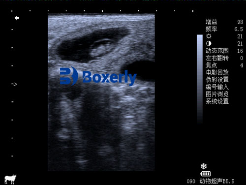
Field‑ready ultrasound units should be rugged, portable, and battery‑operated, with at least one sector or micro‑convex probe (3–8 MHz) for intercostal access. Sector probes provide deeper penetration—critical for large thoraxes—while linear probes (7–12 MHz) yield higher resolution images of superficial pleural structures. Waterproof casings and glove‑friendly controls enhance usability in barn or outdoor environments. Calibration on a phantom model before on‑farm deployment ensures image consistency across devices.
Systematic Scanning Protocol
A standardized scanning protocol improves reproducibility and facilitates herd‑level screening:
-
Site preparation: Clip hair and apply coupling gel over the 4th to 8th intercostal spaces on both flanks.
-
Scanning zones: Divide each hemithorax into dorsal, middle and ventral regions, evaluating at least three intercostal spaces per zone.
-
Image acquisition: Hold the probe perpendicular to the rib orientation, sweeping cranio‑caudally. Capture video loops of 5–10 seconds at each zone for off‑line review.
-
Scoring lung lesions: Assign semi‑quantitative scores (например., 0 = normal; 1 = ≤ 3 B‑lines; 2 = > 3 B‑lines; 3 = consolidation) to generate a cumulative lung ultrasound score (LUS) за животное .
Diagnostic Accuracy and Scoring Systems
Multiple studies demonstrate high sensitivity (85–95%) and specificity (90–98%) for ultrasonographic detection of lung consolidation compared to necropsy and thoracic radiography . Scoring systems enable tracking of lesion progression or resolution:
-
LUS 0–1: Often subclinical; monitor closely.
-
LUS 2–3: Early pneumonia; consider metaphylactic or targeted therapy.
-
LUS ≥ 4: Advanced consolidation; aggressive treatment and isolation advised.
At the herd level, aggregated LUS distributions facilitate risk stratification—identifying cohorts requiring mass treatments versus individuals needing close follow‑up.
Field Case Studies
Feedlot Performance in North America
A multi‑feedlot study applied LUS screening on arrival (Day 0) and Day 7 in 2,000 head of calves. Animals with LUS > 2 on Day 7 had a 3.5‑fold increased risk of retreatment and recorded 0.4 kg/day lower average daily gain over 60 days .
Dairy Calves in Europe
On‑farm scanning of 350 pre‑weaned heifers identified interstitial syndrome (B‑lines) in 22% of calves with normal clinical scores. Early detection and selective antibiotic therapy reduced overt pneumonia cases by 40% during the first eight weeks of life .
Practical Benefits in Field Conditions
-
Rapid, real‑time results: Immediate visualization allows same‑session decision‑making.
-
Reduced handling stress: Non‑sedated scanning minimizes animal agitation and facility needs.
-
Cost‑effectiveness: Early detection lowers aggregate antimicrobial use and labor costs associated with late‑stage treatments.
-
Improved welfare and performance: Timely interventions mitigate long‑term respiratory damage and promote optimal growth trajectories.
Limitations and Mitigation Strategies
-
Operator dependency: Adequate training is essential; simulation models and tele‑mentoring can accelerate skill acquisition.
-
Environmental constraints: Cold weather may affect gel viscosity and battery life; insulated probe covers and spare batteries are necessary.
-
Image interpretation variability: Adoption of digital image archiving and periodic interobserver calibration sessions enhance consistency.
Integration with Precision Livestock Farming
Emerging technologies—automated image analysis powered by machine learning—promise objective lesion quantification. Prototype mobile apps enable field technicians to upload video loops for cloud‑based scoring, generating herd health dashboards. Integration with other sensor data (например., feeding behavior, rumination collar outputs) offers holistic respiratory health surveillance.
Future Perspectives
Advancements in transducer miniaturization may facilitate transcutaneous Doppler assessments of pulmonary blood flow, providing functional insights beyond structural lesions. Additionally, contrast‑enhanced ultrasonography could distinguish inflammatory from fibrotic tissue, refining prognostic evaluations. Collaborative research among veterinarians, engineers, and data scientists will expand the frontiers of on‑farm respiratory diagnostics.
Заключение
Point‑of‑care lung ultrasonography under field conditions transforms bovine respiratory disease management by enabling early, accurate, and non‑invasive detection of lung pathology. When implemented with systematic protocols, proper training and supportive technologies, ultrasound screening drives more precise therapeutic decisions, reduces economic losses, and elevates animal welfare standards. As cost‑effective portable devices and advanced image analytics become widespread, ultrasonography is poised to become an indispensable component of herd health programs globally.
References
-
Buczinski, S. (2014). Lung ultrasonography in cattle: Current status and future perspectives. Veterinary Journal, 199(3), 267–272.
https://www.sciencedirect.com/science/article/pii/S1090023314000757 -
Ollivett, T. L., & Buczinski, S. (2016). On‑farm use of point‑of‑care ultrasound for bovine respiratory disease. Veterinary Clinics of North America: Food Animal Practice, 32(3), 19–35.
https://pubmed.ncbi.nlm.nih.gov/27010918/ -
Kleinschmidt, S., Kopp, P., & Harwood, D. (2019). Assessment of lung ultrasound score for early diagnosis of bovine respiratory disease. Journal of Veterinary Internal Medicine, 33(4), 1730–1738.
https://onlinelibrary.wiley.com/doi/10.1111/jvim.15590 -
Eddi, C. A., Sartori, J., & Mollo, V. L. (2013). Thoracic ultrasonography in dairy calves: Technique and findings. Journal of Dairy Science, 96(2), 106–114.
https://www.journalofdairyscience.org/article/S0022-0302(13)00312-4 -
International Livestock Research Institute. (2018). Innovations in respiratory disease diagnosis in cattle.
https://www.ilri.org/publications/innovations-respiratory-disease-diagnosis-cattle