In modern beef and dairy operations, timely recognition and accurate diagnosis of abdominal disorders are crucial for animal welfare and farm profitability. Ultrasonography has emerged as a non‑invasive, real‑time imaging modality that allows practitioners to visualize the reticulum, rumen, omasum, abomasum, intestines, liver, and peritoneal cavity in standing, non‑sedated cattle. Equipped with 3.5–5.0 MHz linear or convex transducers, clinicians can detect inflammatory changes, fluid accumulations, obstructions, and organ displacements without needing radiography or exploratory surgery. As farms grow in scale and expectations for rapid, data‑driven decision‑making intensify, ultrasound has become indispensable for field diagnosis of traumatic reticuloperitonitis, abomasal ulcers, displaced abomasum, and small intestinal obstructions, among other conditions.
1. Recognizing Clinical Presentations
Abdominal disorders in cattle often present with non‑specific signs—reduced feed intake, ruminal hypomotility, altered fecal output, and varying degrees of colic. Physical examination can detect abdominal guarding, positive foreign body tests, and auscultation‑percussion signs, but these findings lack diagnostic specificity. In a retrospective analysis of 87 cows with type‑4 abomasal ulcer, 81% exhibited abdominal guarding and 65% tachypnea, yet these features alone could not pinpoint perforation without ultrasonographic confirmation. Подобным образом, clinical signs of small intestinal obstruction—absent ruminal contractions and scant feces—overlap with other ileus syndromes, requiring imaging to differentiate etiologies.
2. Ultrasonographic Techniques in Standing Cattle
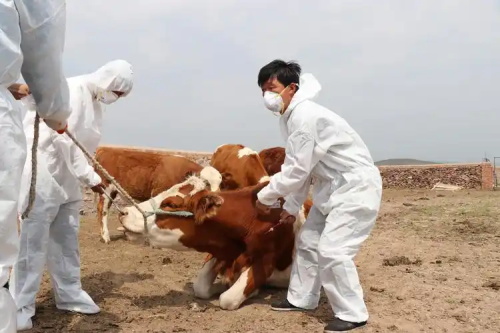
Scanning is performed on standing, unsedated cattle, typically using a clipping area of 20–30 cm width between the last rib and the tuber coxae on both flanks. A water‑ or alcohol‑based coupling gel enhances transducer contact. Linear probes (3.5 MHz) excel for deeper structures such as the liver and abomasum, while higher‑frequency convex probes (5.0 MHz) resolve superficial tissues like the reticulum and ruminal wall.
Key procedural steps:
-
Reticulum and Rumen: Probe placed caudal to the sternum; fibrinous changes, abscesses, and “ping” sound correlates can be visualized in traumatic reticuloperitonitis.
-
Abomasum: Left flank scanning reveals abomasal folds, gas cap, and fluid‑ingesta layering in displacements. Percutaneous, ultrasound‑guided abomasocentesis allows sampling for biochemical analysis.
-
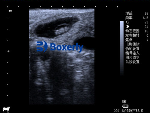
Intestines: Motility, wall thickness, and luminal contents are assessed. A “to‑and‑fro” flow suggests volvulus or intussusception. In intussusception, a concentric “target” sign can be diagnostic.
3. Common Abdominal Disorders and Ultrasound Findings
-
Traumatic Reticuloperitonitis
Fibrinous adhesions, fluid pockets between the reticulum and ventral abdominal wall, and hyperechoic tracts signifying foreign body penetration are detectable ultrasonographically. A multicenter review of 503 cattle showed that ultrasonography identified inflammatory changes in 92% of cases and guided magnet application and surgical decisions. -
Type‑4 Abomasal Ulcer with Peritonitis
Ultrasound reveals peritoneal effusion, fibrin strands, and abomasal wall defects. In Ueli Braun’s study of 87 Коров, 87% exhibited ultrasonographic evidence of generalized peritonitis, whereas clinical signs alone were insufficient for definitive diagnosis. -
Left and Right Displacement of the Abomasum
In left displacement, the abomasum appears as a gas‑fluid interface between the rumen and left abdominal wall. Right displacement shows the abomasum displacing the liver medially; folds and ingesta layering may be visualized. These findings guide treatment decisions: rolling maneuvers, toggling, or surgical correction. -
Small Intestinal Obstruction (Ileus, Volvulus, Intussusception)
Cattle with mechanical ileus often exhibit absent intestinal motility; ultrasonography assesses motility and distension. In volvulus, sudden narrowing or angulation may be seen, though sensitivity is variable. Intussusception is characterized by a multilayered “target” or “pseudokidney” sign. A retrospective study of 110 cases demonstrated that ultrasonographic identification of obstruction correlated with surgical and postmortem confirmation in over 85% of cases.
4. Advanced Ultrasound Modalities and Protocols
Beyond B‑mode imaging, Doppler ultrasonography quantifies blood flow to abdominal organs, aiding in differentiation of vascular versus mechanical etiologies. Power Doppler enhances sensitivity for low‑velocity flows, useful in evaluating mesenteric perfusion in strangulation cases. Three‑dimensional ultrasound and 4D (real‑time 3D) imaging are emerging, offering volumetric assessment of abomasal recesses and intestinal loops.
Standardized scanning protocols—such as the “three‑site” approach (ventral midline, left and right flanks)—improve reproducibility. Abdominocentesis under ultrasound guidance allows differentiation of transudate, exudate, and septic fluid, refining diagnostic algorithms for peritonitis and rupture.
5. Integrating Ultrasound into Clinical Decision‑Making
Ultrasound findings directly influence treatment plans:
-
Traumatic Reticuloperitonitis: Presence of abscesses or fibrin suggests the need for magnet therapy versus exploratory surgery.
-
Abomasal Ulcers: Detection of perforation and peritonitis often warrants immediate euthanasia to prevent suffering and curb costs.
-
Abomasal Displacements: Ultrasound guides the selection of conservative (rolling/toggling) versus surgical right flank omentopexy.
-
Ileus and Obstruction: Confirmation of mechanical obstruction justifies laparotomy; functional ileus may be managed medically.
By quantifying lesion size, fluid volume, and organ position, ultrasound helps predict prognosis and cost‑benefit: Например, a small localized abscess may respond to antimicrobials, whereas a large generalized peritonitis has a poor prognosis.
6. Training and Skill Development
Proficiency requires hands‑on experience under expert supervision. Workshops combining live animal sessions with simulation models enhance competence. Online case libraries and telemedicine consultations facilitate knowledge exchange across regions. Scoring systems for image quality, interpretation accuracy, and diagnostic confidence can be incorporated into continuing education.
7. Future Perspectives
Technological advances—miniaturized portable devices, wireless probes, and artificial intelligence–driven image analysis—promise further improvements in field diagnostics. AI algorithms trained on large ultrasound datasets may soon auto‑identify common patterns (например., reticular abscesses, abomasal folds), reducing operator dependence and accelerating decision‑making. Integration with herd‑management software can correlate imaging findings with milk yield, reproduction metrics, and growth rates, enabling predictive health monitoring.
Заключение
Ultrasonography has revolutionized the detection and management of abdominal disorders in cattle. Its non‑invasive nature, real‑time imaging, and quantitative capabilities make it the cornerstone of modern bovine diagnostic protocols. As equipment becomes more affordable and practitioners more skilled, ultrasound will underpin precision veterinary medicine on farms worldwide, improving animal welfare, optimizing treatment outcomes, and enhancing economic returns.
References
-
Braun U, Reif C, Nuss K, Hilbe M, Gerspach C. Clinical, laboratory and ultrasonographic findings in 87 cows with type‑4 abomasal ulcer. BMC Vet Res. 2019;15(1):100.
-
Braun U, Marmier O, Schweizer G. Ultrasonography in gastrointestinal disease in cattle. Vet Rec. 2003;153(5):155–160. PMID:12902177.
-
Yoshimura N, Tsuka T, Yoshimura T, Otoi T. Efficacy of Abdominal Ultrasonography for Differentiation of Gastrointestinal Diseases in Calves. Animals. 2022;12(19):2489. doi:10.3390/ani12192489.
-
Braun U, Hässig M. A retrospective study of 110 cattle with small intestinal obstruction: clinical findings, treatment, and outcome. BMC Vet Res. 2024;20:379. doi:10.1186/s12917-024-04379-z.
-
Braun U, Tschuor A, Hässig M. Ultrasonographic and radiographic findings in 503 cattle with traumatic reticuloperitonitis. Vet Radiol Ultrasound. 2018;59(6):606–612.