For today’s pig producers, understanding the reproductive physiology of sows is critical not only for optimizing fertility but also for improving productivity and economic return. One of the most important aspects of sow reproduction is follicular development, a dynamic and hormonally regulated process that determines the timing of ovulation and the success of breeding.
With the help of ultrasonography—particularly real-time B-mode ultrasound—farmers and veterinarians can now visualize and monitor ovarian activity in a safe, non-invasive manner. This technological advancement has become especially valuable in both commercial pig farms and artificial insemination centers around the world.
В этой статье, we’ll explore the entire follicular development process in sows, interpret it through the lens of foreign practices and veterinary science, and explain how ultrasonography is revolutionizing how we manage breeding schedules, hormone synchronization, and sow fertility.
Understanding Follicular Development in Sows
The sow’s reproductive cycle is composed of four phases: proestrus, течка, metestrus, and diestrus. Follicular development begins in the proestrus phase and continues until ovulation occurs during течка. On average, a mature sow’s estrous cycle lasts 21 Дни недели, и successful reproduction depends on the timely maturation and ovulation of healthy ovarian follicles.
Key Phases of Follicular Growth
-
Recruitment Phase (Follicular Wave Initiation)
This phase begins a few days after the end of the previous estrous cycle. Small antral follicles (1–2 mm in diameter) begin to grow under the influence of follicle-stimulating hormone (FSH). Dozens of follicles may be recruited, but only a few will proceed to the next phase. -
Selection and Dominance Phase
Among the recruited follicles, several begin to grow more rapidly and become dominant follicles, typically reaching sizes of 4–6 mm. This process is driven by both FSH and luteinizing hormone (LH). The largest follicles suppress the growth of smaller ones through the production of estrogen and inhibin. -
Pre-ovulatory Maturation Phase
The dominant follicles continue growing and reach sizes of 7–10 mm just before ovulation. LH surge triggers final maturation. These follicles develop a thicker wall, increased vascularization, and are ready to release the oocyte. -
Ovulation
Occurs about 36–44 hours after the onset of standing estrus. In a typical multiparous sow, 15–25 follicles will ovulate simultaneously, releasing oocytes that can be fertilized by sperm within the next 8–10 hours.
Follicular Development Under Ultrasonography
The use of B-mode ultrasonography has become common in veterinary practice, especially in Europe, North America, and advanced swine production countries like Denmark and Canada. Portable veterinary ultrasound machines allow practitioners to track follicular development in real-time, improving decisions around breeding and synchronization protocols.
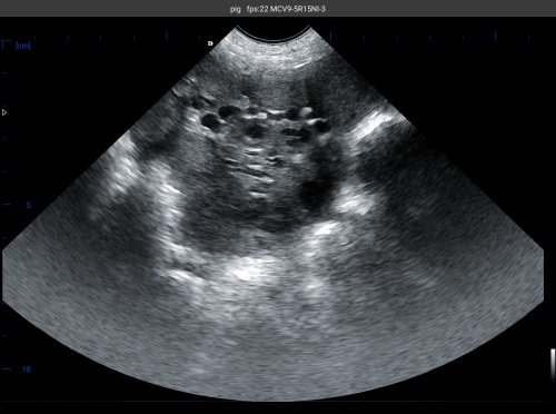
Typical Findings on Ultrasound
-
Small follicles (1–3 mm): Appear as small, round, fluid-filled anechoic (black) structures on the ovary.
-
Dominant follicles (4–6 mm): Become more prominent and begin to crowd out smaller follicles.
-
Pre-ovulatory follicles (7–10 mm): Clearly visible, evenly round, with defined edges.
-
Corpus luteum (CL): After ovulation, the CL appears as a solid, hypoechoic (dark gray) mass, indicating ovulation has occurred.
By using ultrasound, particularly during the late proestrus and estrus periods, farmers can predict the optimal time for insemination, significantly improving fertilization rates.
Hormonal Regulation and Global Practices
In Europe and North America, precise hormonal synchronization programs have been widely adopted. These programs utilize PMSG (Pregnant Mare Serum Gonadotropin) и hCG (human chorionic gonadotropin) to stimulate follicle development and ovulation, especially in gilts or post-weaning sows.
Foreign research, such as that from the University of Alberta and Denmark’s SEGES Pig Research Centre, emphasizes combining ultrasonography with fixed-time AI (FTAI) to minimize handling stress and labor costs. This strategy works by synchronizing all females in a batch, then confirming follicle status using ultrasound before insemination.
In China and Southeast Asia, these practices are being increasingly adopted in larger swine enterprises, though small-scale farmers still rely on behavioral estrus signs, which are less reliable.
Importance of Follicular Monitoring in Problematic Sows
Some sows fail to return to estrus post-weaning or show irregular cycles. In these cases, ultrasound imaging is especially helpful in identifying:
-
Anovulatory follicles (Кисты): Persistently enlarged follicles that fail to ovulate.
-
Delayed follicular growth: Small, underdeveloped follicles even 5–7 days post-weaning.
-
Polycystic ovarian appearance: A sign of chronic reproductive disorder, often linked to endocrine imbalances or over-conditioning.
Foreign literature (например., Soede et al., 2011) highlights the role of body condition, stress, and lighting in influencing follicular waves. Ultrasound helps correlate visible ovarian activity with management conditions.
Follicular Development and Seasonal Effects
Seasonal infertility is a major issue in temperate climates, especially during summer. High temperatures suppress LH pulsatility, reduce estrus expression, and impair follicular growth.
Studies from Spain and the southern United States suggest that ultrasound-guided management—including cooling systems, light control, and dietary modifications (например., high-energy feed pre-weaning)—can help mitigate these effects. Monitoring follicle dynamics with real-time ultrasound allows early intervention and improves summer conception rates.
Practical Benefits of Using Ultrasound on the Farm
From real-world experience and scientific consensus, the following benefits of ultrasound monitoring in sow follicular development are clear:
-
🐖 Increased Breeding Efficiency: Knowing exactly when follicles are mature avoids missed ovulations and increases litter size.
-
🔬 Non-Invasive & Repeatable: No need for blood sampling or invasive procedures. Ultrasound can be done frequently and safely.
-
⏱️ Time-Saving with FTAI: Improves the effectiveness of fixed-time AI protocols, reducing variability.
-
💰 Economic Return: Better timing means higher farrowing rates, larger litters, and more pigs weaned per sow per year.
-
📈 Data-Driven Breeding Decisions: Allows selection of gilts or sows with consistent and optimal follicular response.
Заключение: A Technology-Driven Future in Swine Reproduction
Тем follicular development process in sows is not just a biological phenomenon—it is a management opportunity. As swine production systems become more sophisticated, using ultrasound to monitor ovarian activity will become as standard as using scales or automated feeders.
By understanding the stages of follicular development and integrating real-time imaging, swine farmers around the world can boost fertility, reduce breeding failures, and maximize sow productivity. This is especially true in large-scale operations, where even small improvements in breeding efficiency can translate into thousands of dollars in additional revenue.
Whether you’re managing a 50-sow backyard farm or a 5,000-head commercial operation, ultrasonography offers a clearer window into your sows’ reproductive status—and the tools to act on it.
Reference Sources:
-
Soede, N. M., Langendijk, P., & Kemp, B. (2011). Follicle development and ovulation in sows. Reproduction in Domestic Animals, 46(Suppl 2), 57–63. https://doi.org/10.1111/j.1439-0531.2011.01854.x
-
Knox, R. V. (2016). The use of ultrasound in determining the reproductive status of sows. Theriogenology, 85(9), 1531–1539. https://doi.org/10.1016/j.theriogenology.2015.10.012
-
SEGES Pig Research Centre (2020). Reproductive management using ultrasound. https://pigresearchcentre.dk
-
University of Alberta – Swine Reproduction Lab. https://www.ualberta.ca/afns/swine-reproduction/index.html
-
Whitaker, D. A., & Smith, E. (2021). Veterinary Ultrasonography in Food-Producing Animals. Journal of Veterinary Imaging.