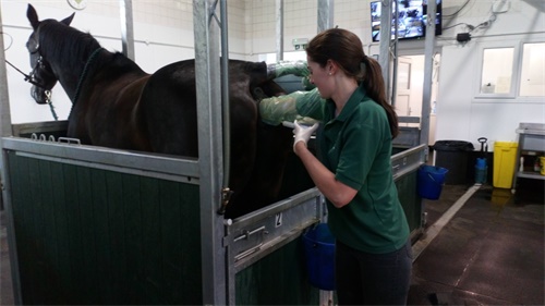
As an equine veterinarian or reproduction specialist, understanding and monitoring the health of pregnant mares is critical—not only for the welfare of the mare but also for ensuring a successful breeding program. One of the most effective, non-invasive technologies available is ultrasound imaging. This article discusses how we—veterinary ultrasound technicians—employ detailed ultrasonographic assessment to evaluate placental health, with a special focus on Combined Thickness of Uterus and Placenta (CTUP), amniotic and allantoic fluid analysis, fetal heart and activity, and hormonal biomarkers. By combining ultrasound findings with endocrine data, we aim to identify early indicators of placentitis, placental insufficiency, or fetal stress, enabling timely intervention. This method aligns closely with international standards of reproductive management in horses.
1. Ultrasonographic Assessment of Placental Health
1.1 Understanding CTUP Measurements
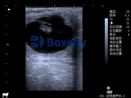
The CTUP—combined thickness of uterus and placenta—measured via transrectal ultrasonography in the caudal chorioallantoic region adjacent to the cervical star, is the most widely accepted quantitative marker for detecting placental pathology like ascending placentitis .
-
Normal reference values vary by gestation stage and breed, but general thresholds are:
-
< 8 mm up to day 300, < 10 mm up to day 330, и < 12 mm thereafter .
-
Draft breeds or large Warmbloods may present slightly higher “normal” CTUPs of up to 15 mm .
-
Early research by Troedsson & Renaudin established these normative ranges and linked increases above two standard deviations after day 271 with placental insufficiency and potential abortion .
1.2 Identifying Placental Separation and Fluid Echoes
Ultrasound scanning can also reveal placental separation or the presence of hyperechoic (bright) particles in fetal fluids, indicative of infection or hemorrhage .
-
Echogenic amniotic or allantoic fluid may signal inflammatory debris, meconium passage, or early placental breakdown .
-
In transabdominal scans, identifying localized thickening—especially around the cervical star—can aid in detecting nocardioform placentitis or hematogenous infections .
1.3 Evaluating Fetal Well‑Being
A comprehensive evaluation also includes fetal heart rate and movement.
-
Normally, fetal heart rates range between ~120–130 bpm early in gestation and decline slightly as term approaches .
-
Tachycardia, bradycardia, reduced activity, or persistent irregularities may signal fetal distress .
Real-time ultrasound enables evaluation of fetal movements, position, and any signs of entanglement or malposition.
2. Integration with Hormonal and Biomarker Data
2.1 Endocrine Indicators of Feto‑Placental Health
Ultrasound findings become far more informative when combined with endocrine monitoring. Key hormones include:
-
Progestagens: Normally moderate until a rise 15–21 days pre-foaling. An early spike may indicate stress or inflammation; a sudden drop is strongly associated with impending abortion .
-
Estrogens (например., estradiol-17β): Levels between 150–280 days above 1,000 pg/ml support normal fetal development; significantly lower levels suggest placentitis or fetal compromise .
-
Relaxin: Lower-than-normal blood levels reflect placental dysfunction, though its use is limited by variable availability .
2.2 Acute Phase Proteins
Serum amyloid A (SAA) can rise sharply in inflammatory conditions like placentitis; however, its specificity is poor—it may elevate due to other systemic infections—so it should be used alongside ultrasound and hormonal assays .
3. Detection and Management of Placentitis
3.1 Clinical Signs to Watch
Early signs of placentitis are typically subtle but may include:
-
Premature udder development and lactation (prepartum lactation) .
-
Vulvar or vaginal discharge suggesting infection due to ascending bacteria .
Physical signs should prompt immediate ultrasound examinations of CTUP and fluid conditions.
3.2 Timeliness Matters
Placental disease, especially ascending placentitis, may progress rapidly—often in late gestation (eight to ten months) . There’s a critical window: the earlier ultrasonographic changes are identified (thickened CTUP, fluid abnormalities), the greater the chance for effective intervention and delivery of a viable foal .
4. Veterinary Intervention Protocols
4.1 Diagnostic Sampling
Alongside imaging, clinicians should collect:
-
Vaginal or cervical swabs for bacteriology and antibiogram.
-
Serial hormone assays (progesterone, estrogen).
-
Acute-phase biomarkers like SAA.
-
Ideally, histopathology of the placenta post-foaling or in case of abortion to confirm ascending placentitis .
4.2 Therapeutic Strategy
A combined approach is usually most effective:
-
Antibiotic therapy: Typically broad-spectrum; agents like potentiated sulfonamides or penicillins are preferred because of their ability to cross into allantoic fluid .
-
Anti‑inflammatories: NSAIDs (например., flunixin meglumine), and methylxanthines like pentoxifylline to reduce inflammation and improve placental circulation .
-
Progestagen supplementation: Oral altrenogest (например., Regumate®) supports uterine quiescence and may suppress preterm labor; double dosing is sometimes used .
-
Adjunctive care: Oxygen therapy during foaling in cases of placenta previa (red-bag delivery), and neonatal antibiotic prophylaxis for foals at risk .
4.3 Monitoring and Outcome Prediction
After initiating therapy, weekly or biweekly ultrasounds reassess CTUP and fetal condition. A return toward normal thickness suggests positive response; persistent thickening indicates need to adjust treatment .
Clinical reports show treatment can yield up to 80–83% live foal outcomes versus 0% in untreated cases .
5. Case Study: A Successful Intervention
A 14‑year-old Warmblood mare—mammary filling observed at day 304—had CTUP elevated above reference by transrectal ultrasound. Immediate therapy began using oral altrenogest, trimethoprim-sulfamethoxazole, flunixin meglumine, and estradiol based on culture results .
Milk calcium and pH were monitored; mammary tone fluctuated during early treatment. By day 331, the mare foaled a viable but small foal; placenta exhibited pus pockets consistent with subclinical placentitis but fetal survival was attributed to early detection and intervention .

6. International Standards and Best Practices
Veterinarians in Europe, North America, and Australia utilize transrectal and transabdominal ultrasound—combined with hormonal monitoring—for routine late-gestation surveillance .
-
In Ireland and the UK, descending CTUP thresholds correspond closely to North American values .
-
Draft breeds, Warmbloods, and Thoroughbreds vary slightly in normative values, emphasizing the need for breed-specific references .
Ultrasound remains the cornerstone tool due to its safety, immediacy, and quantitative capabilities, paired with endocrine assays for a robust, data-driven evaluation.
Заключение
Accurate, early detection of placental abnormalities in pregnant mares is essential for ensuring high fertility and reproductive success. Veterinary technicians and veterinarians should integrate routine late-gestation ultrasound—particularly CTUP measurement—with fluid analysis, fetal monitoring, and biomarker testing. When placentitis is detected early, a combined regimen including antibiotics, NSAIDs, progestagens, and frequent ultrasounds can dramatically enhance the likelihood of a live, healthy foal. As illustrated in international breeding programs, this comprehensive, evidence-based approach represents best practice in modern equine reproductive management.
References
-
Troedsson MHT, Renaudin CD. Transrectal Ultrasonography of the Placenta in Normal Mares and Mares with Pending Abortion, Proc. AAEP Annual Convention, 1997.
-
Renaudin C, Troedsson MH, Macpherson M. Monitoring placental thickness: CTUP reference ranges, Pferdeheilkunde, 2011.
-
LeBlanc MM, Macpherson M. Ascending Placentitis: Pathophysiology, Diagnosis, Treatment, AAEP Proceedings, 2004.
-
Duggan V. Ascending placentitis: A review, PMC, 2008.
-
Kimura K et al., Pregnancy monitoring in mares: Ultrasonographic and endocrine perspectives, Equine Vet J, 2023.
-
Acta Vet Scand (2009).
-
Vet Times (Crabtree, J.), Uses of late-term placental and fetal ultrasound in mares, 2012.
-
ScienceDirect (2020). Estradiol 17β and progestogen as biomarkers.