For horse breeders, veterinarians, and equine specialists, ensuring the health of a full-term foal before birth is critical to optimizing survival rates and long-term performance. The final stages of fetal development—particularly organ maturation—are decisive for the newborn’s ability to adapt immediately after delivery. With the rise of advanced veterinary technologies, real-time ultrasound scanning has become an indispensable, non-invasive tool for monitoring the organ development of full-term foals, allowing for timely interventions when necessary.

În acest articol, we will explore how real-time ultrasound technology is used to assess the maturation of vital organs in full-term foals, why this method is gaining traction globally, and how its practical applications are transforming equine breeding management.
Understanding Organ Maturation in Fullterm Foals
The final trimester of equine gestation is a period of rapid growth and complex organ development. While the skeletal and muscular systems have largely formed earlier, the last stages of gestation focus on:
-
Lung maturation (critical for respiration)
-
Liver function establishment
-
Kidney development
-
Gastrointestinal tract preparation
-
Cardiac and circulatory refinement
These developments are essential for the foal to transition from intrauterine life—where oxygen and nutrients are supplied via the placenta—to independent postnatal existence. Failure of any organ system to mature adequately can lead to neonatal maladjustment syndrome, respiratory distress, or multi-organ failure after birth.
Historically, assessing fetal well-being during late pregnancy relied heavily on maternal signs, hormonal assays, or indirect indicators such as fetal movements. Însă, these methods offered limited insights into the actual state of fetal organs. This is where real-time ultrasound scanning provides a major breakthrough.
How Real-Time Ultrasound Scanning Works for Foal Organ Monitoring
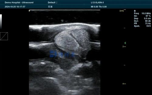
Ultrasound imaging employs high-frequency sound waves to generate real-time images of internal fetal structures without exposing the mare or fetus to ionizing radiation. With Modul B (brightness mode) ultrasonography, veterinarians can obtain detailed cross-sectional images of fetal organs, observing size, formă, and structural integrity.
More advanced systems may also integrate Doppler ultrasound to assess blood flow, allowing evaluation of placental function and fetal circulation—critical components in assessing fetal health status.
Key scanning windows during full-term gestation include:
-
Thoracic cavity for lung and cardiac assessment
-
Abdominal cavity for liver, kidney, and gastrointestinal observation
-
Umbilical cord and placenta to evaluate vascular flow and placental sufficiency
Through these real-time images, veterinarians can monitor whether organ development is proceeding normally, or whether there are signs of delayed maturation that may necessitate intervention or specialized neonatal care post-delivery.
The Global Veterinary Perspective on Fullterm Foal Monitoring
In countries with strong equine industries—such as the United States, the United Kingdom, Australia, and Germany—the use of real-time ultrasound in late-term gestation monitoring is increasingly considered standard practice. Leading veterinary associations and equine reproductive specialists emphasize several reasons for this:
1. Non-Invasive and Safe for Repeated Use
Unlike certain biochemical tests that require invasive sampling, real-time ultrasound poses no risk to the fetus or the mare, even with repeated examinations. This allows for regular monitoring of at-risk pregnancies.
2. Early Detection of Complications
Fetal anomalies, delayed organ development, or placental insufficiencies can be identified before they result in stillbirth or neonatal morbidity. Early detection enables veterinarians to prepare for assisted deliveries or prompt interventions.
3. Improved Decision-Making
Breeders can make informed choices about timing of delivery, including whether induction might be necessary if the mare’s condition deteriorates while the foal’s lungs are still underdeveloped.
4. Enhanced Neonatal Preparedness
If underdeveloped organs are identified, neonatal specialists can prepare life-saving interventions such as supplemental oxygen, surfactant therapy, or intensive care for weak foals immediately after birth.
Organ-Specific Ultrasound Evaluation at Full Term
Let’s examine how real-time ultrasound specifically evaluates major organ systems in the full-term foal:
Lung Maturation
The maturation of pulmonary tissues is perhaps the most critical factor in full-term foal viability. On ultrasound, lung tissue transitions from a relatively anechoic (fluid-filled) appearance earlier in gestation to a more echogenic, structured appearance as alveoli form and mature.
Studies in equine reproduction show that correlating ultrasound lung appearance with biochemical markers of surfactant production can accurately predict pulmonary maturity. Without mature lungs, the foal is at high risk for respiratory distress syndrome at birth.
Cardiac Development
Real-time ultrasound allows detailed visualization of cardiac chambers, valve function, and even measurement of fetal heart rate variability. Abnormalities such as arrhythmias or structural defects can be detected, providing critical information for delivery planning.
Liver and Kidney Development
Liver size and echotexture can be assessed to estimate metabolic readiness, while kidney development is evaluated based on corticomedullary differentiation. Abnormal organ sizes may signal intrauterine growth retardation or impending metabolic complications postpartum.
Placental and Umbilical Blood Flow
Through Doppler ultrasound, blood flow through the umbilical arteries and veins, as well as uterine arteries, can be analyzed. Reduced placental perfusion may suggest placental insufficiency, a leading cause of fetal growth restriction and stillbirth.
Real-Life Application: A Case Study Approach
On a thoroughbred breeding farm in Kentucky, veterinarians employed real-time ultrasound monitoring on a 345-day pregnant mare with a history of prior stillbirths. Initial scans revealed underdeveloped lung echogenicity and marginal umbilical artery flow. With daily monitoring, clinicians observed progressive improvement in lung appearance.
At 355 Zile, with favorable ultrasound findings, the delivery was carefully supervised, and the foal required minimal respiratory support at birth. The ability to track fetal organ maturity with precision ultimately contributed to a successful outcome.
Ultrasound-Guided Interventions in Equine Perinatology
As real-time ultrasound adoption grows, veterinarians are developing protocols for ultrasound-guided decision-making:
-
Delay elective induction if lungs remain immature
-
Administer corticosteroids to stimulate fetal lung maturation in high-risk pregnancies
-
Prepare neonatal ICU resources preemptively when organ immaturity is detected
-
Conduct assisted deliveries with surgical or pharmacological support based on predicted neonatal weakness
Such strategies highlight how real-time ultrasound is not merely diagnostic but becoming central to equine perinatology management.
Challenges and Limitations
Despite its tremendous advantages, real-time ultrasound monitoring is not without challenges:
-
Operator Skill: Accurate interpretation requires significant training and experience.
-
Equipment Cost: High-resolution ultrasound units capable of equine fetal imaging are costly, limiting access for smaller breeders.
-
Foal Positioning: Fetal positioning late in gestation may obscure certain organs from optimal imaging windows.
-
Limited Normative Data: Research is still ongoing to establish universal ultrasound reference standards for equine fetal organ maturity.
Ongoing international collaboration among veterinary colleges and breeding associations is helping to address these gaps.
The Future of Real-Time Ultrasound in Foal Monitoring
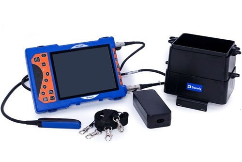
With advances in imaging resolution, portable veterinary ultrasound devices, such as the latest models available on the market, continue to improve accessibility. Innovations like 3D/4D ultrasound and artificial intelligence-assisted image interpretation promise even greater accuracy in assessing fetal organ health.
As more breeders globally recognize the life-saving potential of prenatal ultrasound, its role will likely become as routine as other standard prenatal care protocols in equine medicine.
Concluzie
The last weeks of gestation represent a critical window for foal survival, where even subtle delays in organ maturation can have life-threatening consequences. Real-time ultrasound scanning empowers veterinarians and breeders to visualize, monitor, and act upon fetal development with unprecedented precision.
By providing real-time insights into lung readiness, cardiac function, liver health, and placental perfusion, ultrasound enables proactive decision-making that dramatically improves neonatal outcomes. Its adoption across equine breeding programs worldwide is not just a technological trend—it represents a fundamental advancement in animal welfare and reproductive success.
As technology continues to evolve and become more accessible, real-time ultrasound will undoubtedly become an indispensable part of every serious equine breeder’s management toolkit.
Reference Sources:
-
Renaudin, C., et al. (2022). “Ultrasound Imaging in Equine Fetal Monitoring: Recent Advances and Clinical Applications.” Equine Veterinary Journal, 54(3), 321-330. https://beva.onlinelibrary.wiley.com/journal/20423306
-
Johnson, Un. L., & Giguère, S. (2020). “Neonatal Foal Medicine: Assessment of Fetal Readiness for Birth.” Veterinary Clinics of North America: Equine Practice, 36(2), 273-290. https://www.sciencedirect.com/journal/veterinary-clinics-of-north-america-equine-practice
-
Fowden, Un. L., & Forhead, Un. J. (2015). “Endocrine Mechanisms of Fetal Maturation in the Horse.” Equine Veterinary Journal, 47(5), 527-534. https://beva.onlinelibrary.wiley.com/doi/full/10.1111/evj.12376