In modern swine production, the ability to accurately assess breeding traits in live animals is essential for improving productivity, meat quality, and economic returns. Among the diagnostic tools used in this pursuit, veterinary ultrasonography has gained considerable traction. Although widely adopted in clinical and production settings, especially in North America and Europe, the application of ultrasound in pig breeding still faces several technical and practical challenges.
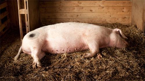
Veterinary ultrasonography, particularly real-time B-mode imaging, offers a non-invasive way to evaluate internal body structures such as fat thickness, muscle area, and even intramuscular fat (IMF) content. In countries like the United States, the National Swine Improvement Federation (NSIF) has established certification standards for technicians using ultrasound in live pig evaluation. This has accelerated the integration of ultrasonography into genetic improvement programs. Însă, its broader adoption—especially in Asia and developing markets—is still limited by issues like accuracy, device portability, image interpretation, and cost-effectiveness.
This article examines how veterinary ultrasonography is currently being used in pig breeding programs, what technical challenges remain, and how the global community is approaching these challenges through research, standardization, and innovation.
Ultrasound in Pig Breeding: A Global Perspective
Veterinary ultrasonography in pig breeding is primarily used to evaluate phenotypic traits that are otherwise difficult or impossible to assess in live animals. These include:
-
Loin muscle depth or area (often the Longissimus dorsi)
-
Intramuscular fat (IMF) Niveluri
-
Reproductive organ assessment
In the United States and parts of Europe, these parameters are routinely measured in selection candidates to guide breeding decisions. The ability to predict carcass quality and reproductive potential without slaughtering the animal allows for more sustainable and data-driven breeding programs. Despite this, international studies continue to emphasize that image quality, operator experience, and equipment variability can greatly affect the precision of these measurements.
Sources of Measurement Error in Pig Ultrasonography
Several factors limit the accuracy and consistency of ultrasonographic readings in pigs. These can be broadly categorized into:
-
Image Acquisition Challenges
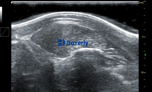
The accuracy of ultrasonic predictions is significantly influenced by the breed of pig, the anatomical structure being measured, and the physiological status of the animal. Variables such as fat and muscle depth, age, sex, and animal movement during scanning can all impact the results. Unlike cattle or sheep, pigs often do not remain still during scanning, leading to misaligned probe positioning and distorted images.
-
Subjectivity in Image Interpretation
Another major limitation is the subjectivity involved in interpreting the ultrasonic images. Even trained technicians may vary in their assessments of the same scan, especially in complex evaluations such as IMF content. This subjectivity is further compounded by the limited number of cross-sectional slices typically obtained during scanning. Since these slices provide only 2D snapshots of a 3D body, they may not fully capture the complete tissue distribution.
-
Equipment Constraints
Portable ultrasound devices used on farms must be durable enough to withstand harsh conditions—humidity, temperature fluctuations, dust, and animal waste. Many devices currently on the market may lack sufficient resolution or sensitivity to detect subtle differences in tissue structure. Moreover, regular maintenance and calibration are needed to ensure consistent performance, which can be a financial burden for small-scale producers.
International Solutions and Best Practices
Several countries have tackled these limitations by implementing protocols, training programs, and technology upgrades. De exemplu:
-
In the U.S., technicians are certified through the NSIF, which ensures a standard level of competency in acquiring and interpreting ultrasonic images.
-
European breeding organizations often follow harmonized protocols that dictate probe placement, image acquisition timing, and data storage formats.
-
Advanced algorithms and artificial intelligence (AI) are increasingly being used to assist in image interpretation, reducing operator bias and improving reproducibility.
Efforts are also underway to integrate 3D imaging and advanced software modeling into routine evaluations. These enhancements could help reconstruct more complete representations of muscle and fat distribution in pigs, providing more precise input for breeding value estimation.
Interpretation Standards and Data Consistency
For ultrasonic data to be meaningful across different herds, farms, and even countries, clear measurement protocols are essential. For pigs, this includes:
-
Standardized body locations for scanning (typically between the 10th and 11th ribs)
-
Defined minimum and maximum weight or age ranges during measurement
-
Replicated measurements to ensure consistency
-
Calibration of probes and software with reference carcass data
Foreign experts have emphasized the importance of recording environmental and management variables alongside ultrasonic data. Differences in diet, housing, health status, and handling procedures can introduce variability into measurements, and these factors must be accounted for when interpreting results or comparing them across groups.
Ultrasound in IMF Prediction: The Holy Grail?
Among the most challenging applications of veterinary ultrasonography in pig breeding is the prediction of intramuscular fat, also known as marbling. IMF plays a key role in meat tenderness and flavor, making it an essential quality trait, particularly in premium markets such as Japan and South Korea.
Research, especially in the United States, suggests that real-time ultrasound has the potential to non-invasively estimate IMF content with reasonable accuracy. Improvements in image resolution and post-processing software now allow technicians to detect differences in muscle echo patterns that correlate with marbling levels.
Însă, these applications are still under development and have not yet been widely adopted in commercial pig production due to cost and complexity. Nevertheless, the future is promising. As both software and imaging hardware continue to evolve, IMF prediction could soon become a routine component of genetic selection programs.
Health and Biosecurity Considerations
Another often-overlooked aspect of applying ultrasound in live pig evaluation is disease control. Probes used on multiple animals or across herds can become vectors for pathogen transmission if not properly disinfected. International protocols recommend using species-specific disinfectants and requiring operators to follow strict hygiene practices, especially in facilities housing high-value breeding stock.
Global pig breeding programs, particularly those in disease-sensitive regions like the European Union, incorporate biosecurity as a non-negotiable aspect of ultrasound use. This includes having dedicated scanning rooms, single-use covers, and UV-based sterilization systems.
Cost and Feasibility in Farm Management
For ultrasonography to be useful at the commercial level, especially in developing countries, it must be economically viable and easy to implement in routine management. Foreign experts stress that:
-
The cost of ultrasound devices must be within the budget range of average producers.
-
Operators must receive adequate training to avoid generating misleading or useless data.
-
The time required per scan should not disrupt regular farm operations.
Lightweight, battery-powered devices with user-friendly interfaces and cloud-based data storage options are making headway in markets such as Brazil, Vietnam, and China. Paired with mobile apps and remote technician support, these innovations are helping democratize access to advanced breeding tools.
The Road Ahead: Integration with Genomic Selection
Veterinary ultrasonography is becoming an important partner in genomic selection. By pairing phenotypic data from ultrasound scans with genomic profiles, breeders can more accurately estimate breeding values and make faster genetic gains.
De exemplu, a pig showing favorable ultrasonic traits—such as deep loin muscle and low backfat thickness—can be confirmed as genetically superior using single-nucleotide polymorphism (SNP) testing. Conversely, discrepancies between genotype and phenotype can highlight measurement errors or suggest previously unknown genetic factors.
The integration of ultrasound data into genomic databases is already standard practice in some breeding companies in Europe and the U.S., and this trend is expected to expand globally.
Concluzie
Veterinary ultrasonography holds immense promise in transforming pig breeding from an experience-based art into a data-driven science. Despite current limitations in accuracy, subjectivity, and equipment cost, ongoing international efforts in training, standardization, and technological development are addressing these issues.
The use of ultrasonography in breeding programs not only improves selection accuracy but also enhances animal welfare by reducing the need for invasive assessments. As research continues to improve the technology’s predictive power—especially in evaluating intramuscular fat—ultrasound is poised to become an indispensable tool in the genetic improvement of pigs worldwide.
By learning from best practices abroad and tailoring them to local conditions, producers and researchers can unlock the full potential of this powerful technology.