In modern cattle reproduction management, precision is paramount. Whether optimizing conception rates or understanding ovarian health, ultrassonografia veterinária has become an indispensable diagnostic tool. Among its many applications, one particularly valuable use is the monitoring of follicular development in naturally cycling cows. While the general process of folliculogenesis is well understood, less attention has been paid to the potential differences in follicle development between the left and right ovaries. Recent observations using veterinary ultrasound suggest there may be subtle asymmetries in how follicles grow, mature, and regress in each ovary. Understanding these differences can improve reproductive decision-making and shed light on bovine reproductive physiology.
Veterinary Ultrasound in Ovarian Monitoring
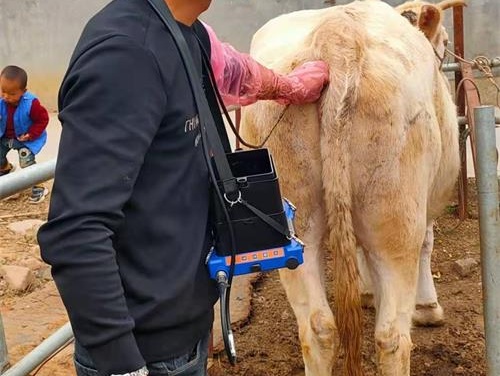
Ultrassom veterinário, specifically B-mode imaging, is a non-invasive, real-time tool that allows veterinarians and researchers to visualize internal reproductive structures in live animals. When applied to the ovaries, this imaging method excels at identifying and tracking the development of follicles, corpus luteum, and cysts.
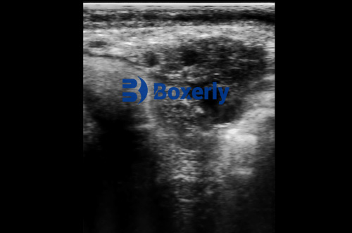
Follicles appear as round, fluid-filled, anechoic (black) regions on the monitor. O follicular wall and surrounding ovarian tissue reflect the ultrasound waves and appear as echogenic (white or gray) areas. As follicles grow, their shape may range from perfectly round to slightly oval or irregular, especially in response to pressure or spatial constraints within the ovary.
In contrast, the corpus luteum (CL) appears as a grayish, moderately echogenic structure. During early luteal development, it may contain a small central cavity, appearing as a hypoechoic region within the CL.
The Natural Estrous Cycle and Follicle Development
In cows, the estrous cycle typically lasts about 21 Dias. Veterinary ultrasound enables daily monitoring of this cycle, revealing consistent patterns of follicular emergence, selection, dominance, and ovulation.
Here’s a generalized timeline of key events during the bovine estrous cycle:
-
Day 0–1: Ovulation occurs. A dominant follicle ruptures, releasing the oocyte.
-
Day 3–4: Multiple small follicles begin to appear on both ovaries.
-
Day 10: The corpus luteum reaches its maximum size, providing high progesterone secretion.
-
Day 15: Luteolysis begins; the CL regresses.
-
Day 18–20: A new dominant follicle emerges, leading to the next ovulation.
Ultrasound allows for real-time observation of these changes. Por exemplo, a dominant follicle will appear as a large, black circle with a thin wall, while a regressing CL becomes smaller and less echogenic.

Comparing Follicle Development in Left vs. Right Ovaries
While cows have two ovaries, they are not always equally active. Various studies and field observations suggest a slight but noticeable difference in follicle development between the left and right ovaries.
In a study involving young heifers monitored by veterinary ultrasound, the following average values were recorded:
-
Left Ovary:
-
Dominant follicle diameter: approximately 11.85 milímetro
-
Number of visible follicles: around 3.18
-
-
Right Ovary:
-
Dominant follicle diameter: approximately 13.09 milímetro
-
Number of visible follicles: around 3.09
-
These differences are small and generally not statistically significant. Contudo, they consistently indicate that the right ovary may slightly outperform the left in follicular activity.
Furthermore, during the ovulatory wave, the average diameter of the pre-ovulatory follicle was:
-
Left Ovary: sobre 14.76 milímetro
-
Right Ovary: sobre 14.34 milímetro
Again, the difference is minimal, suggesting relatively balanced development between sides. Contudo, in practice, veterinarians and breeders have observed a higher incidence of pregnancy establishment in the right uterine horn. This suggests that while follicular development may be symmetrical, the uterine environment or ovulation efficiency may differ.
Potential Causes of Ovarian Asymmetry
The reasons for asymmetry in ovarian function are not fully understood, but several theories exist:
-
Anatomical Variation
The cow’s right ovary is anatomically closer to the rumen, which may affect blood flow, hormone exposure, or spatial freedom for follicular expansion. -
Vascular Differences
Studies in reproductive physiology have shown that blood flow in the ovarian artery may differ between sides. Since follicle development is dependent on hormonal and nutritional support via the bloodstream, even small differences can influence outcomes. -
Neurological Influence
The innervation of reproductive organs might also play a role. Neurotransmitters and local reflexes could affect follicular recruitment or ovulation differently on each side. -
Hormonal Microenvironment
Although systemic hormone levels are generally balanced, local concentrations of prostaglandins, estrogens, and growth factors may vary between ovaries due to tissue sensitivity or feedback loops.
Clinical and Reproductive Implications
For veterinary practitioners and cattle breeders, the practical question is: Do these slight differences matter?
In most cases, no immediate clinical action is required based on ovary asymmetry alone. Contudo, in cases of repeat breeding or unexplained infertility, veterinary ultrasound assessments of both ovaries can provide critical clues. If one ovary consistently fails to produce mature follicles or the CL regresses prematurely, treatment strategies may be adjusted accordingly.
Adicionalmente, monitoring ovulation side can help with predicting which uterine horn to target for embryo transfer, especially in programs involving synchronized breeding or artificial insemination.
Ultrasound-Based Strategies in Herd Management
Integrating veterinary ultrasound into reproductive management allows for tailored approaches. Por exemplo:
-
Ovulation synchronization protocols (por exemplo,, Ovsynch) can be monitored to ensure timely follicle development.
-
Follicular cysts or persistent follicles can be detected early and treated.
-
Postpartum ovarian recovery can be tracked to assess readiness for breeding.
In herds with intensive reproductive programs, monitoring ovulation patterns on both ovaries helps balance conception rates and improve genetic outcomes by maximizing the use of high-value semen.
Global Perspectives and International Research
Research conducted in North America, Europe, and Australia supports the utility of ultrasound in assessing bovine ovarian health. While follicular symmetry is often assumed, fieldwork confirms that monitoring both sides adds value.
In a 2022 Canadian study, ultrasound monitoring over multiple estrous cycles revealed that the right ovary accounted for 56% of observed ovulations. Similarly, European dairy farms have reported improved reproductive efficiency when ovulation patterns are considered in breeding schedules.
Conclusão
Veterinary ultrasound offers a window into the dynamic world of bovine ovarian physiology. Although the differences between the left and right ovaries in follicular development are subtle, they provide valuable insights for both scientific understanding and practical herd management. By using ultrasound to track follicle growth, veterinarians and breeders can make more informed decisions, optimize breeding timing, and improve pregnancy outcomes.
As this technology becomes increasingly available on farms worldwide, recognizing and interpreting the nuances of ovarian function—on both sides—will continue to play a key role in advancing bovine saúde reprodutiva.