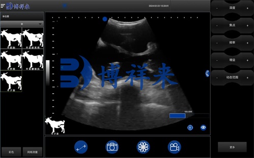
Ultrasound technology has rapidly become a cornerstone of modern livestock management, especially in the reproductive programs of sheep farming. Among its various applications, one of the most valuable—yet often overlooked—is its role in ensuring economic efficiency in embryo transfer (ET) programs. In countries with developed sheep breeding systems such as Australia, New Zealand, and parts of Europe, ultrasound scanning is now a routine part of selecting donor and recipient ewes, monitoring superovulation, and evaluating embryo development and pregnancy success. This article explores how the use of ultrassonografia veterinária can significantly reduce economic losses and increase success rates in sheep embryo transfer, drawing on international practices and research.

Ultrasound as a Screening Tool for Donors and Recipients
In embryo transfer programs, selecting healthy donor and recipient ewes is essential. Contudo, identifying reproductive abnormalities or early pregnancies in animals sourced from external farms can be challenging through visual inspection alone. This is where ultrasonography becomes indispensable.
Using real-time B-mode ultrasound, veterinarians can quickly and accurately assess the reproductive tract. Non-pregnant, cyclic ewes with healthy uterine horns and ovaries are ideal candidates for ET programs. When ultrasound is used to pre-screen animals, already-pregnant ewes or those with uterine abnormalities can be excluded before hormone treatments begin. This prevents:
-
Hormone-induced abortions in undetected pregnancies
-
Wasted medications on unsuitable animals
-
Unnecessary logistical costs in managing non-productive animals
Internationally, many breeding operations in Australia and the UK now use portable, farm-ready ultrasound scanners to perform on-site evaluations, which allow rapid sorting of suitable donor and recipient sheep, reducing unnecessary treatment costs.
Reducing Losses Through Superovulation Monitoring
In typical ET programs, donor ewes undergo superovulation to increase the number of transferable embryos. Traditionally, ovulation responses were only assessed during laparotomy, where visual inspection of the ovaries would be used to count corpora lutea (CLs). This method is invasive and risky.
Modern practices in Canada and France now rely on pre-operative ultrasonographic monitoring. With a linear-array rectal probe, vets can evaluate the ovarian surface and follicular development days before the surgical procedure. This enables:
-
Accurate estimation of expected ovulation numbers
-
Identification of underperforming ewes (por exemplo,, fewer than four CLs)
-
Decision to skip surgery in suboptimal cases, allowing natural mating instead
By selectively excluding low-responding donors, breeders can avoid unnecessary surgery, minimize animal stress, and reduce the risk of complications such as oviduct adhesions—an often irreversible consequence of surgical embryo flushing.
Improving Success Rates Through Timely Embryo Transfer
The success of embryo transfer in sheep depends on several interconnected factors: estrus synchronization, recipient readiness, pregnancy establishment, and fetal retention. Ultrasound contributes at nearly every stage.
In international practice, synchronization records are combined with ultrasound findings to fine-tune the recipient-donor match. Proper timing ensures that recipients are at the exact physiological stage to accept an embryo, significantly boosting pregnancy rates.
After embryo transfer, traditional methods like observing estrus return or teaser ram exposure are often used to detect non-pregnant ewes. Contudo, these methods can miss subtle signs or depend heavily on subjective observations.
Ultrasound offers an objective, early, and accurate solution.

Early Pregnancy Detection and Fetal Counting
Around 21 days post-transfer, a skilled technician can perform transrectal ultrasonography using a high-frequency linear probe to identify early pregnancy indicators such as:
-
Fluid-filled gestational sacs (anechoic zones)
-
Embryonic poles or fetal heartbeat (by 25–30 days)
-
Number of embryos (by assessing discrete sacs)
By 35 Dias, fetal number can often be confirmed with high confidence. This information enables:
-
Grouping pregnant ewes by fetal number
-
Adjusting feed rations to match gestational demands
-
Avoiding overfeeding or underfeeding
-
Prioritizing care for multiple pregnancies, which carry a higher risk
Studies from the University of Guelph in Canada show that grouping and feeding pregnant ewes according to their fetal count improves lamb survival and reduces metabolic stress on the dam.
Embryo Development Monitoring: A Preventive Strategy
Even after a pregnancy is confirmed, ultrasound continues to play a vital role in monitoring the health and development of the embryo. Periodic scanning can detect:
-
Irregular fetal growth
-
Abnormal uterine fluid levels
-
Signs of fetal demise
In regions like Western Europe, where sheep production often uses intensive systems, this level of monitoring is essential to prevent economic losses from unnoticed pregnancy failures. If abnormalities are spotted early, treatment interventions—such as progesterone supplementation or modified nutritional plans—can be initiated, significantly improving fetal retention rates.
International Perspectives on Economic Benefits
A cost-benefit analysis conducted by the New Zealand Ministry for Primary Industries estimated that including routine ultrasound in an ET program saved approximately 18–25% in costs related to wasted hormones, failed pregnancies, and surgical complications. Similarly, in Australia, many breeders now consider ultrasound an investment rather than an expense, as it enables better donor selection, avoids unnecessary embryo wastage, and allows dynamic herd management.
In Spain, a study published in Veterinaria Clínica reported that farms using ultrasound-based screening prior to ET procedures saw a 12% increase in lambing rates compared to farms relying solely on visual observation and timing records. The direct economic impact was measured not only in improved lamb numbers but also in reduced veterinary interventions and feed misallocation.
Future Outlook: Technology Adoption and Innovation
Ultrasound technology is advancing rapidly. Modern devices offer:
-
Real-time 3D imaging
-
Doppler functions for blood flow analysis
-
Wireless probes for remote use
-
AI integration for automated pregnancy detection
In high-tech farms across the U.S. and Europe, these devices are now being used not just by veterinarians but also by trained farm personnel, reducing labor costs and improving response time. With further miniaturization and affordability, sheep farmers in developing countries will also gain access to these tools, unlocking greater reproductive efficiency globally.
Conclusão
Ultrasound scanning in sheep embryo transfer is no longer a luxury—it is a necessity for any operation serious about maximizing efficiency and profit. From identifying the most suitable donor and recipient ewes, monitoring superovulation responses, and increasing surgical efficiency, to confirming early pregnancy and tracking fetal development, ultrasound enables informed decisions at every stage.
International practices have shown that ultrasound-guided ET programs result in fewer complications, lower costs, and higher success rates. As the technology continues to evolve and become more accessible, its economic and practical benefits will become even more pronounced.
For any sheep producer looking to improve reproductive outcomes and reduce unnecessary expenditure, investing in portable veterinary ultrasound equipment and training is a forward-thinking, cost-effective strategy.
Reference Sources:
-
Peterson, C. G., & Lee, B. H. (2022). “Ultrasound Applications in Small Ruminant Reproduction.” Journal of Animal Reproduction Science.
-
Ministry for Primary Industries, New Zealand (2023). “Economic Evaluation of Embryo Transfer Technology in Sheep Production.”
https://www.mpi.govt.nz/et-ultrasound-economic-evaluation -
Pérez, R. et al. (2021). “The Impact of Early Pregnancy Detection Using Ultrasound in Spanish Sheep Farms.” Veterinaria Clínica, 45(3), 34–41.
https://www.vetclinica.es/sheep-et-ultrasound-impact