Ultrasound is a cornerstone imaging modality in veterinary medicine. Para profissionais de animais de grande porte e operadores de fazendas, it’s an invaluable tool that enhances diagnostic speed and accuracy without the need for radiation. Among the more elusive anatomical structures to locate are the adrenal glands—small but powerful endocrine organs that play a central role in hormone regulation.
Finding these glands consistently on ultrasound can be challenging, especially in species like cattle, swine, e cabras, where body conformation and fat distribution may obscure visibility. This article provides a practical guide on how to reliably identify adrenal glands during veterinary ultrasound exams, tailored especially for field use in large animal practice.
Why Locate the Adrenal Glands?
The adrenal glands produce essential hormones such as cortisol, aldosterone, and adrenaline. Abnormalities in their size, forma, or echotexture can indicate endocrine disorders, Tumores, stress-related pathologies, or systemic illnesses. Early detection is critical for improving animal welfare and productivity, especially in livestock settings where herd health impacts profit margins.
Understanding the Anatomy
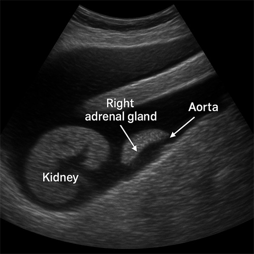
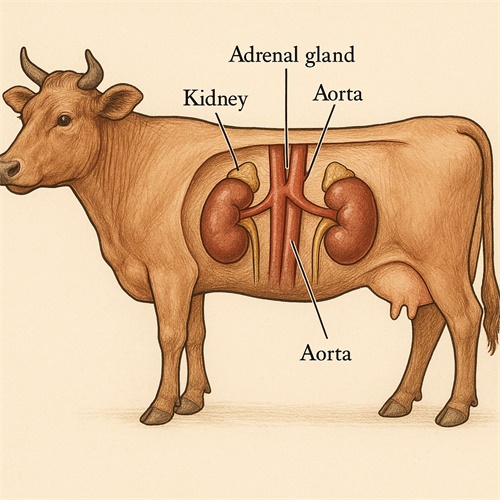
The adrenal glands are typically located cranial and medial to the kidneys. In ruminants like cows, they are positioned just ahead of the cranial pole of the kidney, near the aorta. The right adrenal gland is often harder to image due to its proximity to the caudal vena cava and the gas-filled gastrointestinal tract. In pigs, the glands are more deeply embedded in retroperitoneal fat and may require higher-frequency probes and proper fasting before scanning.
Ultrasound Machine Settings
Before beginning the scan, make sure your ultrasound machine is optimized:
-
Use a microconvex or linear probe (5–8 MHz for adult cattle; 7.5–12 MHz for small ruminants and pigs)
-
Apply ample ultrasound gel
-
Adjust depth to 5–10 cm depending on the animal’s size
-
Turn off harmonics for better tissue differentiation
-
Activate compound imaging if your device supports it
Probe Placement and Scanning Technique
To find the adrenal glands, follow these general steps:
-
Start with the kidney
Begin by locating the kidney in longitudinal view. This acts as your anatomical anchor. In cattle, begin scanning from the right paralumbar fossa to find the right kidney, then move cranially. -
Move cranially and medially
From the cranial pole of the kidney, shift your probe cranially and medially toward the area just lateral to the aorta. This is where the adrenal gland typically lies. -
Adjust angle and pressure
Apply gentle but firm pressure and adjust the angle to avoid gas shadows from the intestine. Slight oblique angles may offer a clearer image. -
Identify key features
The adrenal gland in large animals is often oval or peanut-shaped with a hypoechoic (darker) cortex and a more echogenic (brighter) medulla. It should measure around 2–4 cm in length in adult cattle.
Tips for Different Species
Cattle:
-
Jejum 12 hours before scanning can reduce rumen gas interference.
-
The right adrenal is more accessible than the left due to less ruminal coverage.
-
Use rectal or transabdominal approach depending on age and size.
Goats and Sheep:
-
Due to smaller size, a linear probe with higher frequency (7.5–12 MHz) offers better resolution.
-
The glands are more superficial and can be found near the last rib under the epaxial muscles.
Swine:
-
Deep body fat and gas can make adrenal imaging difficult.
-
Fasting and left lateral recumbency help improve visibility.
-
The glands are usually more hyperechoic compared to cattle.
When Are the Adrenals Abnormal?
Recognizing pathology is as important as identifying normal anatomy. Look out for:
-
Enlargement: May indicate hyperadrenocorticism (Cushing’s syndrome)
-
Irregular margins or nodules: Suggestive of adrenal tumors or metastases
-
Hyperechoic areas: May signal mineralization or chronic degeneration
-
Asymmetry: In bilateral organs, size difference greater than 2:1 can be abnormal
Por exemplo, in dairy herds with sudden milk drop and weight loss in cows, ultrasound examination of the adrenal glands can help rule in or out endocrine issues that might be secondary to chronic stress or neoplasia.
Conclusão
Locating the adrenal glands using veterinary ultrasound may require practice, but it’s a highly rewarding skill. For large animal practitioners and livestock managers, it empowers early diagnosis of significant hormonal disorders. With proper preparation, equipment, and technique, adrenal imaging becomes a practical part of routine health checks and diagnostics on the farm.
References and Further Reading
For further reading and case-based guidance, consult:
-
“Bovine Adrenal Ultrasonography: Techniques and Pitfalls” by University of Florida CVM
https://extension.vetmed.ufl.edu -
“Ultrasonographic Anatomy of the Normal and Diseased Adrenal Glands in Small Ruminants” – Journal of Veterinary Diagnostic Investigation
https://journals.sagepub.com/home/vdi -
“Large Animal Diagnostic Imaging” – Veterinary Clinics of North America: Food Animal Practice
https://www.sciencedirect.com/journal/veterinary-clinics-of-north-america-food-animal-practice