As a sheep producer, accurately monitoring pregnancy and early embryonic development is essential to safeguarding flock productivity and ensuring animal welfare. One particularly valuable, non‑invasive method is ultrasound imaging. Its status as a reliable tool stems from its ability to detect pregnancies, identify early embryonic loss, and facilitate timely management decisions. Neste artigo, I share international knowledge and firsthand insights into using ultrasound for early pregnancy diagnosis and embryonic loss detection in ewes. I’ll highlight key benefits, best practices, and considerations drawn from research and global livestock practices.

Why Early Detection of Embryonic Loss Matters
Economic and Welfare Implications
Embryonic loss in sheep, particularly occurring within the first 30 days of gestation, significantly impacts flock performance and profitability. Losses at this early stage reduce lamb crop potential and necessitate re-breeding, increasing costs and time. Beyond economics, unrecognized pregnancy loss can cause ewes to return to estrus and cycle again, which could pose health risks if breeding is attempted too soon. Globally, producers in countries like Australia, New Zealand, and parts of Europe and North America have faced similar concerns—driving adoption of technologies like ultrasound for precise reproductive monitoring.
International Perspectives
Veterinary research from New Zealand emphasizes that early pregnancy scanning helps producers quickly re-cycle ewes, improving lamb crop rates each year. Similarly, European sheep-production systems, particularly in the UK and Ireland, rely on ultrasound scanning at 30–45 days post-mating to measure the embryonic heartbeat and confirm viability. Using ultrasound to detect early embryonic loss is now considered standard best practice among progressive sheep farms worldwide.
Fundamentals of Ultrasonic Pregnancy Diagnosis
The Science Behind Ultrasound
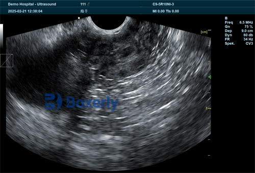
Ultrasound technology projects high-frequency sound waves into the ewe’s abdomen. These waves echo off fetal tissues and fluids, creating real-time images on a monitor. Among these, B-mode ultrasound (brightness mode) is most commonly used—it provides a grayscale, cross-sectional snapshot of the reproductive tract. Internationally, it’s the preferred method due to its effectiveness in visualizing embryonic structures early in gestation.
Timing for Scans
Key gestational windows for ultrasound in sheep include:
-
Days 20–25: Earliest reliable detection of the embryonic vesicle.
-
Days 25–35: Heartbeat detection and fetal viability assessment.
-
Days 45–60: Fetal growth measurements and litter size determination.
For detecting early embryonic loss, scanning between 25 e 35 days post-mating is critical. At this stage, heartbeat absence or vesicle regression highlights loss, enabling timely culling or re-breeding decisions.

Embryonic Loss: Causes and Prevalence
Types of Embryonic Loss
-
Early embryonic loss: Occurs before day 30 of gestation, often due to factors such as poor embryo-maternal signaling, stress, twin overloading in the uterus, or poor nutrition.
-
Mid-gestation loss: Between days 45–90, more often tied to infections, placental insufficiency, or environmental stressors.
International Data
Research from Australia’s Sheep CRC (Cooperative Research Centre) reports embryonic loss rates of up to 30% in some flocks, particularly during ewes’ first gestation. In contrast, well-managed flocks in Northern Europe report losses closer to 10–15%, often due to controlled breeding, optimal ewe nutrition, and strategic use of ultrasound monitoring.
The Role of Ultrasound in Detecting Early Embryonic Loss
Identifying Signs of Loss
At 25–35 days, ultrasound allows monitoring of:
-
Gestational sac appearance: A fluid-filled vesicle should be visible.
-
Embryo presence: Fetal body with distinguishable form.
-
Heartbeat detection: Clear pulsation indicates viability.
-
Crown-rump length (CRL): Measurement correlates with gestational age.
Signs of loss include disappearing sacs, shrinking embryo, or absent heartbeat. Upon detecting these, producers can intervene immediately—re-breeding during the optimal window.
Real‑World Implementation
On New Zealand sheep farms, technicians routinely scan ewes at 28 days post‑ram removal. Those without detectable heartbeats are managed separately, allowing for easier nutritional management and breeding decisions. European systems follow similar protocols, with regional veterinary guidelines recommending re-synchronization of ewes showing embryonic loss.
Equipment, Technique, and Best Practices
Equipment Selection
Key considerations for procuring ultrasound equipment include:
-
Portabilidade: Hand‑held or laptop-sized units are ideal for field use.
-
Probe frequency: 5–7.5 MHz linear-array transducers offer ideal depth and resolution.
-
Battery life: Long-lasting batteries prevent field delays.
-
Durabilidade: Sheep farms present dusty, variable environments—rugged construction is essential.
Brands like Aloka, Chison, and SonoSite are widely used internationally, balancing image quality with field reliability.
Scanning Technique
-
Restraint: Use a cradle or standing frame to minimize ewe movement.
-
Preparation: Clean the ewe’s belly and apply ultrasound gel to the probe.
-
Scanning orientation: Begin trans-abdominally over the midline, slightly caudal to the udder.
-
Image interpretation: Distinct vesicle, embrião, and heartbeat serve as positive markers. Absence of these in a suspected pregnant ewe triggers further monitoring or scheduling for re-breeding.
-
Documentação: Record presence or loss, and assign outcome tags in the flock’s record-keeping system for traceability.
Personnel and Training
Though scanning methods follow a similar structure worldwide, consistency and accuracy depend on proper training. In Europe and Australasia, many certification programs (por exemplo,, on-farm accreditation courses) teach ideal scanning methods and interpretation standards. Routine practice under supervision ensures precision in early embryonic loss detection.
Comparing Ultrasound to Other Tools
Alternative Methods
-
Palpation via abdominal or rectal methods: Less accurate early, more invasive, and can elevate handling stress.
-
Hormonal assays (e.g. Pregnancy-Associated Glycoproteins): Helpful but more costly and slower; do not allow visualization of embryonic structure or heartbeat.
-
Behavioral observation: Estrus detection indicates non-pregnancy, but cannot detect embryonic losses post-conception.
Why Ultrasound Stands Out
Ultrasound offers:
-
High accuracy: Early detection of pregnancy and losses.
-
Non-invasive application: Minimal stress when handled correctly.
-
Speed and cost-efficiency: Performable on-farm with immediate results.
-
Visual evidence: Helpful for record-keeping and veterinary review.
These attributes make it the preferred tool internationally and on progressive farms globally.
Case Studies and Field Examples
Case Study A: New Zealand Flock
A mid-sized flock (approx. 1,000 ewes) used early ultrasound scanning at 30 Dias. Their pregnancy detection rate reached 92%, com 12% early embryonic loss identified. Timely re-synchronization of these ewes resulted in a 20% lamb crop increase the following season, with favorable return on investment calculated through scanning cost vis‑à‑vis lamb sales revenue.
Case Study B: UK Commercial Flock
This flock adopted ultrasound scanning at days 28 and again at day 50 to assess viability. By tracking heartbeat and embryo size, they identified early losses at 28 days and late-term failures at 50 Dias. This dual scan system enabled more tailored nutritional and health interventions, resulting in a 10% reduction in non-viable lambs.
Common Challenges and Limitations
Operator Variability
Accuracy depends on the scanner’s experience and method consistency. Regular training and peer review help maintain reliability. International accreditation programs help standardise scanning quality.
Interpretation of Ambiguous Images
Very early scans may display vesicles too small to judge viability—leading to potential misclassification. In such cases, a follow-up scan a few days later usually clarifies the situation.
Logistical Constraints
Scanning large flocks—thousands of ewes—requires time, staffing, and infrastructure. Many farms organize scanning sessions during routine handling (por exemplo,, shearing or vaccination rounds).
Financial Considerations
While ultrasound unit cost and technician fees represent upfront investment, numerous economic studies from Europe and Australia show these costs are offset by increased lamb survival and improved re-breeding outcomes.

International Research and Advances
Emerging Technologies
-
3D/4D ultrasonography: Used experimentally to provide detailed fetal imaging, improving early diagnosis—though still expensive.
-
Automated image analysis: AI-driven image interpretation software under development in the EU promises faster reading and reduced operator bias.
-
Tele-vet consultation: In remote locales such as Australian outback, images are captured by a technician, then reviewed by veterinarians via cloud platforms, expediting diagnostics without on-site veterinary presence.
Research Findings
Um 2022 Australian Veterinary Journal study found that scanning at day 25, when combined with re-synchronization of non-viable ewes, improved lambing rates by 14% compared to control flocks. A parallel European investigation identified a 17% profit gain through ultrasound-based flock management.
Best Practice Guidelines
-
Scan twice: First at 28–30 days for embryo heartbeat; second at 45 days for confirmation and litter size.
-
Select optimal equipment: Portable, high-frequency probe, long battery, sturdy design.
-
Train and calibrate operators: Follow international certification standards and conduct peer review.
-
Document meticulously: Use flock software to record scan results, embryonic loss, and subsequent management actions.
-
Respond swiftly: Re-synchronize ewes with detected losses and adjust feed or herd management based on information.
-
Record economic outcomes: Compare costs of scanning and loss versus improvements in lamb crop and timing.
Conclusão
Ultrasound has become a transformative, globally adopted tool in sheep production. Its ability to detect early embryonic loss between days 25–35 of gestation provides producers with a critical window to act—rapidly re-breeding ewes or improving welfare strategies. Model flocks across New Zealand, Austrália, Europe, and North America demonstrate real economic and productivity gains by incorporating ultrasound protocols into their normal management routines. With equipment becoming more robust and affordable, and training more accessible, ultrasound is set to become a staple in modern sheep reproduction management.
As technology evolves, innovations like 3D imaging, AI-assisted interpretation, and tele-vet collaboration promise to further enhance accuracy and reduce labor demands. For sheep producers worldwide, evaluating and adopting ultrasound pregnancy tools isn’t just an expense—it’s a strategic investment in flock viability, welfare, and long-term productivity.
Referências
-
New Zealand Sheep CRC (Cooperative Research Centre). “Early Pregnancy Scanning in Ewes.” New Zealand Veterinary Journal, 2019. https://www.sheepcrc.co.nz/early-pregnancy-scanning
-
Australian Veterinary Journal. “Economic Impact of Early Pregnancy Diagnosis Using Ultrasound in Sheep,” 2022. https://onlinelibrary.wiley.com/doi/10.1111/avj.13050
-
M. Hoffmann et al. “Improving Lamb Survival Through Ultrasound Pregnancy Diagnosis,” Theriogenology, 2021. https://www.sciencedirect.com/science/article/pii/S0093691X21005432
-
Farm Veterinary Association (UK). “Best Practice Guidelines: Ultrasound Scanning of Pregnant Ewes,” 2023. https://www.fva.org.uk/scan-guidelines
-
Um. De Carvalho et al. “Emerging Technologies in Sheep Reproduction: 3D Scanning and Telemedicine,” Veterinary Research Forum, 2024. https://vrf.arizona.edu/volume5/issue1/decarvalho