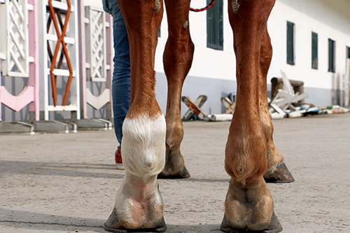
When horses are in training—whether for racing, show jumping, dressage, or general performance—their legs take the brunt of the work. Tendons, ligaments, joints, and bones are all vulnerable to injury, especially with repeated strain. On horse training farms, even small undetected limb injuries can escalate into serious, career-ending problems if not caught early. That’s where ultrasound has quietly become a game-changer.
Veterinarians and trainers across the world are now relying on portable ultrasound machines not just in emergency situations, but as part of their routine monitoring protocol. These devices offer a non-invasive, real-time window into what’s going on inside the equine limb—helping catch injuries long before the horse starts limping.
The Realities of Limb Injuries on Training Farms
Horses in training are athletes. Like human athletes, they face stress injuries, repetitive strain, and the occasional acute trauma. The biggest challenge? Many injuries in their early stages are invisible to the naked eye. A horse might appear sound—moving normally, eating, training—yet be dealing with micro-tears in a tendon or early signs of inflammation in a ligament.
In the U.S., U.K., and Australia, it’s common practice for racehorse trainers to schedule regular imaging during the racing season. Why? Because the cost of losing a horse to a preventable injury is astronomical. Ultrasound plays a central role in this proactive approach. It helps identify slight changes in tissue echogenicity, swelling, and fiber alignment—clues that the leg is under duress.
What Ultrasound Actually Sees
A veterinary-grade ultrasound doesn’t just take pictures; it tells a story. It sends sound waves into the tissue, which bounce back and create an image. With a trained eye, vets can spot subtle disruptions in the pattern of soft tissue—especially in tendons and ligaments.
Common structures imaged include:
-
Superficial digital flexor tendon (SDFT)
-
Deep digital flexor tendon (DDFT)
-
Suspensory ligament
-
Check ligament
-
Joint capsules and synovial sheaths
Changes in echogenicity (brightness on the screen) can indicate inflammation, fiber damage, or fluid build-up. Over time, ultrasound also lets vets track healing progress, comparing new scans to previous ones to determine how the injury is responding to treatment.
Preventative Scanning: A Global Shift
Horse owners in Europe and North America are increasingly asking for regular ultrasound exams—not just when an issue arises. One popular method is to perform baseline scans of tendons and ligaments at the beginning of a training season, then compare these scans at regular intervals.
In the U.K., por exemplo, many Thoroughbred training facilities have integrated biweekly limb ultrasounds into their management plan. It’s viewed as a form of “predictive maintenance”—catching the silent signs of overuse before they turn into tears or ruptures.
Similarly, sport horse barns in Germany and the Netherlands have embraced routine ultrasound, especially for jumping and dressage horses that place significant strain on their suspensory ligaments. Vets there report that having a complete imaging history often makes the difference between a minor rest period and a 12-month rehab.
Ultrassom portátil: More Than Just Convenient
Modern portable ultrasound machines have totally changed the game for equine vets. Compact and rugged, these devices can be brought directly into the barn aisle, paddock, or trailer. And because they provide instant feedback, decisions can be made on the spot.
Por exemplo, if a horse suddenly presents with mild swelling after a workout, a quick scan can determine whether it’s superficial edema or a deeper tendon injury. This immediate clarity often prevents overreacting or, worse, underreacting.
In Australia, many rural equine vets rely on portable ultrasound not just for tendons, but for hoof-related injuries too. Abscess tracking, navicular bursa evaluation, and even some early laminitis changes can be detected using these tools in the field.
Benefits Beyond Detection
Ultrasound is more than just a diagnostic tool—it’s also a roadmap for rehabilitation. Once an injury is detected, the recovery process becomes a highly visual, measurable journey. Vets can document the degree of lesion healing, the quality of fiber realignment, and the presence of scar tissue.
Owners appreciate being able to see the healing themselves. Trainers use these images to pace training resumption carefully, reducing the risk of reinjury. And horses benefit from treatments that are tailored to their individual recovery timelines, not just generic rest periods.
The Learning Curve: Skill Matters
It’s worth noting that equine limb ultrasound isn’t something just anyone can do. It requires training, experiência, and a deep understanding of limb anatomy. That’s why many horse owners work with veterinarians who specialize in imaging or have pursued advanced ultrasound certification.
In the U.S., the American Association of Equine Practitioners (AAEP) regularly hosts ultrasound training workshops. Vets from all over North America attend to sharpen their skills and stay updated on the latest imaging protocols.
Meanwhile, some larger training facilities in Europe have an in-house vet or technician who handles routine imaging, while external specialists are brought in for more complex scans or second opinions.
Ultrasound in Action: Real Farm Experiences
Take, por exemplo, a performance horse stable in Kentucky. They began scanning all their horses monthly during show season. Within a year, they had detected and treated six minor tendon lesions that would otherwise have gone unnoticed until lameness developed.
In Newmarket, a major racing stable used regular ultrasound monitoring to fine-tune their training schedule. When one promising colt showed subtle suspensory changes on a scan, they pulled him from races for two weeks. He returned stronger and won his next two starts.
These stories are becoming more common across the globe. From Argentina to Ireland, trainers are finding that adding ultrasound to the toolkit isn’t just smart medicine—it’s smart business.
Limitations and Responsible Use
While ultrasound is incredibly valuable, it’s not a magic wand. It can’t image bone structure as well as radiography or detect metabolic disorders. And without proper interpretation, it can lead to false assumptions.
That’s why it’s important to use ultrasound alongside other diagnostic tools like flexion tests, radiographs, and clinical assessments. It’s one piece of the puzzle—albeit a powerful one.
Em alguns casos, over-reliance on ultrasound might lead to unnecessary rest or even over-treatment. The key is to pair technology with veterinary experience and good horsemanship.
The Bottom Line
Ultrasound has become an essential tool for detecting limb injuries in horses, especially those in high-demand training environments. It’s safe, não invasivo, rápido, and incredibly informative. As more horse farms across the world embrace regular imaging as part of their management, the result is fewer injuries, shorter downtimes, and better overall outcomes.
Whether you’re managing racehorses, sport horses, or ranch horses, ultrasound helps ensure that every step they take is as safe as possible. In the world of horse training, where every limb matters, this tool is proving to be worth its weight in gold.