In modern equine practice, maintaining optimal reproductive health is one of the most important challenges for breeders, veterinarians, and horse owners. Ensuring the successful development of a healthy fetus is critical not only for the well-being of the mare and foal but also for the economic sustainability of the breeding operation. Among the diagnostic tools available, ultrasonography has emerged as an indispensable, non-invasive technology for identifying fetal abnormalities early in gestation, allowing for informed decision-making and timely intervention. Neste artigo, we will explore how veterinarians apply animal ultrasound scanners to detect fetal abnormalities in horses, examine common types of anomalies, discuss the advantages of ultrasound technology, and highlight how its role continues to evolve in equine reproductive management.

Understanding Fetal Development in Horses
A thorough understanding of normal fetal development is essential for detecting abnormalities. In mares, pregnancy typically lasts about 340 Dias, though variations between 320 e 370 days are not uncommon. Fetal development can be divided into distinct stages:
-
Embryonic Phase (Days 0–45): The embryo develops rapidly, with organogenesis occurring during this period.
-
Fetal Phase (Days 46–Term): Organs mature, body growth accelerates, and fetal structures become increasingly visible on ultrasound.
During these stages, any disruption in growth or organ formation may lead to congenital abnormalities. Detecting these conditions early allows for better management of both the mare and the pregnancy.
The Role of Ultrasound in Equine Pregnancy Monitoring
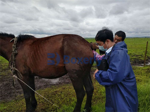
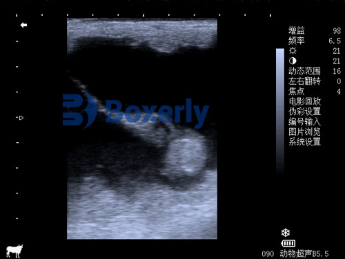
In equine reproduction, ultrasonography is typically performed using transrectal or transabdominal probes, depending on the gestational age and the anatomical area being assessed. The versatility of ultrasound scanners allows veterinarians to visualize fetal development in real time, assess placental health, monitor amniotic fluid levels, and identify potential abnormalities.
-
Transrectal Ultrasound: Most commonly used during early pregnancy (up to 120 Dias), enabling close imaging of the fetus and placenta.
-
Transabdominal Ultrasound: Employed after mid-gestation when the fetus moves into the abdominal cavity and grows beyond the reach of transrectal probes.
The non-invasive nature of ultrasound makes it ideal for repeated monitoring throughout pregnancy without causing discomfort or harm to the mare or fetus.
Common Fetal Abnormalities Detected by Ultrasound
Several fetal abnormalities can be identified via ultrasonography in horses. Early detection plays a vital role in determining the prognosis of the pregnancy and potential outcomes.
1. Hydrocephalus
One of the most recognizable congenital abnormalities is hydrocephalus—an excessive accumulation of cerebrospinal fluid in the brain. Ultrasound imaging can reveal an abnormally enlarged cranium and dilated ventricles, often visible by mid-gestation. Severe cases may result in dystocia (difficult birth), requiring cesarean section or elective termination of the pregnancy.
2. Twin Pregnancies
While not a malformation per se, twin pregnancies pose significant risks in equine reproduction, often leading to late-term abortions or birth complications. Ultrasound allows for early identification of twins, typically around days 14–16 of gestation, enabling veterinarians to manually reduce one embryo to improve the chance of a successful singleton pregnancy.
3. Umbilical Cord Abnormalities
The umbilical cord is a vital lifeline connecting the fetus to the placenta. Ultrasound can detect cord torsions or excessive coiling, which may compromise blood flow and oxygen delivery, potentially leading to fetal death. Regular monitoring helps veterinarians assess the cord’s structure and function as pregnancy progresses.
4. Placental Insufficiency
Placental health is crucial for fetal development. Ultrasonography enables veterinarians to measure placental thickness, evaluate placental attachments, and monitor for signs of placentitis (inflammation of the placenta). Abnormal thickening or detachment of the placenta may indicate compromised fetal nourishment, increasing the risk of abortion or premature delivery.
5. Fetal Growth Retardation
By measuring fetal biometric parameters such as crown-rump length, biparietal diameter, and limb length, veterinarians can assess whether the fetus is growing appropriately for its gestational age. Deviations from normal growth patterns may signal chromosomal abnormalities, Infeção, or poor placental function.
Advantages of Using Ultrasound in Equine Fetal Monitoring
Ultrasound scanning provides multiple benefits that make it the gold standard for fetal monitoring in horses:
-
Imagens em tempo real: Allows dynamic visualization of fetal movement, heartbeat, organ development, and placental function.
-
Não invasivo e seguro: Repeated use is safe for both mare and fetus, eliminating the risks associated with invasive procedures.
-
Early Diagnosis: Abnormalities can be identified weeks or even months before clinical signs would otherwise become apparent.
-
Guidance for Management: Early detection allows for intervention strategies such as medical treatment, nutritional support, or decisions regarding pregnancy continuation.
As equine veterinarians often emphasize, “Early detection is key to early intervention.”
Technical Considerations and Skill Requirements
While ultrasound is a powerful diagnostic tool, its effectiveness relies heavily on the skill of the operator and the quality of the equipment. The following factors are crucial:
-
Operator Expertise: Accurate interpretation requires specialized training and experience in equine reproductive ultrasonography. Misinterpretation of images can lead to unnecessary interventions or missed diagnoses.
-
Equipment Quality: High-resolution scanners, such as B-mode ultrasound systems, provide clearer images, allowing for finer detail in fetal structures. Modern portable ultrasound devices offer advanced features, including Doppler imaging, which assesses blood flow within the umbilical cord and placenta.
-
Timing and Frequency: Routine ultrasound exams are typically scheduled throughout pregnancy, with critical assessments occurring at key gestational milestones:
-
Day 14–16: Confirmação de gravidez, twin check
-
Day 25–30: Fetal heartbeat detection
-
Day 60–70: Organ development review
-
Day 150+: Placental evaluation and fetal growth assessment
-
Case Studies: Practical Applications in Equine Clinics
Case Study 1: Early Twin Reduction
A 12-year-old mare presented for routine breeding management. At 15 days gestation, ultrasound revealed two viable embryos. The veterinarian promptly performed manual reduction of one embryo, allowing the remaining singleton to develop normally. Regular ultrasound follow-ups confirmed healthy fetal growth, and the mare delivered a healthy foal at term.
Case Study 2: Detection of Hydrocephalus
In another case, a mare at 220 days gestation underwent routine ultrasound. The fetus exhibited an abnormally large head with dilated brain ventricles, indicative of hydrocephalus. Due to the likelihood of dystocia, the veterinary team prepared for a planned cesarean section, successfully delivering the foal while minimizing risks to the mare.
Case Study 3: Umbilical Cord Torsion
A 9-year-old mare showed signs of intermittent colic during late gestation. Transabdominal ultrasound revealed excessive umbilical cord torsion with compromised blood flow. Early diagnosis allowed the team to monitor the mare closely and prepare for early intervention. Unfortunately, the foal was delivered prematurely and did not survive, but the mare recovered fully—a positive outcome for future breeding.
Challenges and Limitations of Ultrasound in Equine Practice
Despite its advantages, ultrasound is not without limitations:
-
Interpretation Variability: Operator-dependent skill levels can affect diagnostic accuracy.
-
Equipment Cost: High-quality ultrasound machines can represent a significant financial investment for smaller practices.
-
Limited Reach in Late Pregnancy: As the fetus grows, its position may limit transrectal accessibility. Transabdominal scanning requires experience and optimal probe positioning.
-
Incomplete Diagnosis: Some genetic or functional abnormalities may not be visible on ultrasound alone, necessitating adjunct diagnostics such as genetic testing or advanced imaging.
Nonetheless, ultrasound remains the most practical, accessible, and widely used tool for routine fetal assessment.
The Future of Ultrasound in Equine Reproduction
Technological advancements continue to improve ultrasound Capacidades. Modern systems now incorporate:
-
3D and 4D Imaging: Offering three-dimensional reconstructions and real-time motion analysis.
-
Elastography: Measuring tissue stiffness to assess placental health.
-
Advanced Doppler Techniques: Providing more detailed analysis of fetal and placental blood flow.
Such innovations promise even earlier detection of subtle abnormalities and better understanding of fetal physiology. As these technologies become more portable and affordable, more breeders and equine practitioners worldwide are expected to integrate them into routine care.
Conclusão
The application of animal ultrasound scanners in equine fetal abnormality diagnosis has revolutionized reproductive veterinary practice. By enabling early detection of structural and functional anomalies, ultrasonography supports informed decision-making that ultimately protects both animal welfare and economic viability.
As equine breeders and veterinarians increasingly rely on this technology, its role will only grow in importance, driven by advances in image quality, diagnostic precision, and operator training. The ongoing evolution of ultrasound ensures that equine reproductive management will continue to become safer, more effective, and more responsive to the needs of mares, foals, and the industry as a whole.
Reference Sources:
-
Ginther, O. J. (2014). Ultrasound Imaging and Animal Reproduction: Cavalos. Equiservices Publishing.
-
McCue, P. M., & Ferris, R. Um. (2015). Use of Ultrasonography for Monitoring Equine Pregnancy. The Veterinary Clinics of North America: Equine Practice, 31(1), 129-143. https://www.sciencedirect.com/science/article/pii/S0749073914000640
-
Rantanen, N. W. (2020). Equine Pregnancy Ultrasound. American Association of Equine Practitioners (AAEP). https://aaep.org/proceedings/equine-pregnancy-ultrasound