 BXL
Corpus Luteum Ultrasound in Cattle: How to Examine Ovarian Structures and Improve Reproductive Management
Introduction: Why Is Corpus Luteum Ultrasound Important in Cattle?
Reproductive efficiency is one of the most important factors influencing profitability in modern cattle farming. Whether managing a dairy herd, beef cattle operation, or breeding farm, accurate evaluation of ovarian structures plays a critical role in reproductive management.
Corpus luteum ultrasound is an important diagnostic application in bovine reproductive medicine. By using real-time veterinary ultrasound imaging, veterinarians can examine the ovaries, identify the corpus luteum (CL), evaluate its size and internal structure, and monitor ovarian activity throughout the estrous cycle.
The corpus luteum is a temporary endocrine structure that develops on the ovary after ovulation. Its primary function is to produce progesterone, a hormone essential for maintaining the uterine environment during early pregnancy.
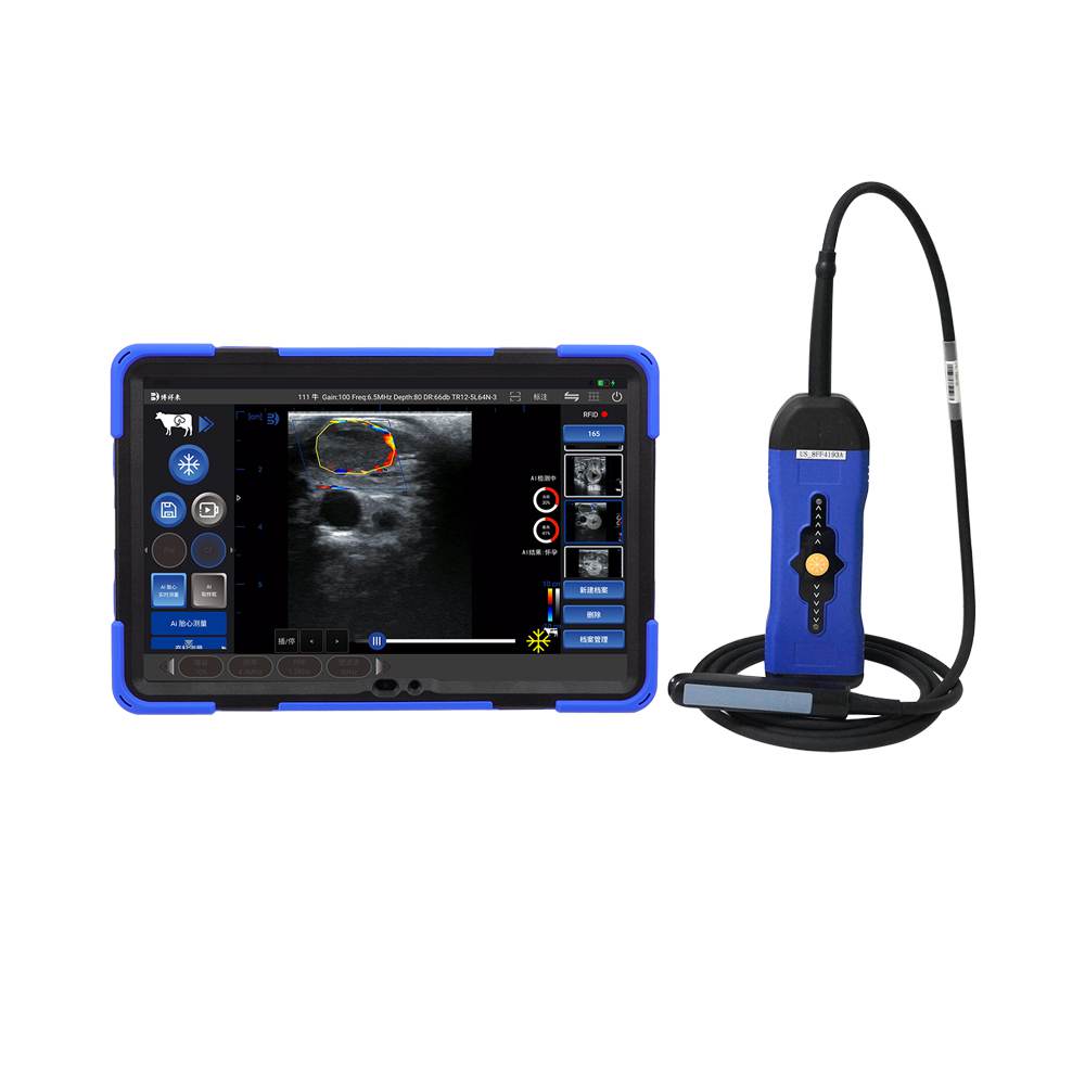
With a suitable veterinary ultrasound scanner, veterinarians can obtain valuable information about ovarian function, reproductive status, and pregnancy-related changes without relying exclusively on manual rectal palpation.
For modern cattle reproduction programs, high-quality ovarian ultrasound imaging provides an effective way to improve diagnostic efficiency and support better breeding decisions.
1. What Is the Corpus Luteum in Cattle?
The corpus luteum, commonly abbreviated as CL, forms from the ovarian follicle after ovulation.
Following the release of an egg, the remaining follicular cells undergo structural and functional changes, developing into the corpus luteum.
Its most important function is progesterone secretion.
Progesterone helps:
– Prepare and maintain the uterine environment for embryo development.
– Support early pregnancy.
– Prevent the development of another ovulatory cycle during pregnancy.
– Regulate the bovine estrous cycle.
In non-pregnant cattle, the corpus luteum normally regresses if pregnancy does not occur. This allows the reproductive cycle to continue.
In pregnant cows, the corpus luteum is generally maintained because progesterone production is essential for pregnancy support.
Portanto, identifying and evaluating the corpus luteum is an important part of bovine ovarian ultrasound examination.
2. How Does Corpus Luteum Ultrasound Work?
9
A veterinary ultrasound scanner uses high-frequency sound waves to produce real-time images of internal reproductive structures.
During a bovine ovarian examination, the veterinarian usually uses a transrectal linear probe to examine the ovaries through the rectal wall.
The ultrasound image can help identify:
Corpus luteum structure
The CL usually appears as a relatively solid ovarian structure with a characteristic echogenicity that differs from surrounding ovarian tissue.
Corpus luteum size
Measuring the CL can help assess its development and changes during the estrous cycle.
Internal structure
Some corpus luteum structures contain a central fluid-filled cavity. This is commonly known as a cavitary corpus luteum. It is a physiological variation and should not automatically be interpreted as a pathological cyst.
Ovarian activity
By examining the CL together with developing follicles, veterinarians can obtain a more complete picture of ovarian function.
High-resolution ultrasound imaging makes these structures easier to distinguish and evaluate.
3. When Should Veterinarians Examine the Corpus Luteum?
Corpus luteum ultrasound is useful at different stages of cattle reproductive management.
Um. During the Estrous Cycle
After ovulation, the corpus luteum develops and increases progesterone production.
Ultrasound examination allows veterinarians to monitor its development and observe changes in ovarian structures.
This information can help evaluate the reproductive stage of individual cows.
B. During Early Pregnancy Diagnosis
The presence of a corpus luteum can provide useful information during early reproductive examinations.
Contudo, the presence of a corpus luteum alone does not confirm pregnancy.
A non-pregnant cow can also have a functional CL during the normal estrous cycle.
For more reliable pregnancy diagnosis, veterinarians should evaluate the uterus, uterine contents, embryonic structures, and other relevant findings.
With advanced veterinary ultrasound equipment, examination of the uterus and ovaries can be performed during the same transrectal scanning procedure.
C. During Reproductive Synchronization Programs
Many modern cattle farms use hormonal synchronization protocols to improve breeding efficiency.
Monitoring ovarian structures can help veterinarians evaluate reproductive responses and make better-informed decisions about insemination timing.