 BXL(ビーエル)
Cattle Embryo Ultrasound: Early Pregnancy Detection and Accurate Embryo Monitoring in Cows
1. What Is Cattle Embryo Ultrasound?
Cattle embryo ultrasound is an advanced diagnostic technique used by veterinarians and cattle reproductive specialists to examine the uterus, identify developing embryos, and monitor early pregnancy in cows and heifers.
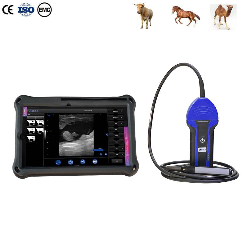
Unlike traditional pregnancy detection methods, ultrasound technology allows the examiner to observe internal reproductive structures in real time. By using a suitable veterinary ultrasound scanner with a specialized rectal probe, veterinarians can identify pregnancy-related changes, visualize the embryo, and evaluate its development.
今日, bovine embryo ultrasound has become an important tool in modern dairy and beef cattle reproduction management.
For dairy farms, beef cattle operations, artificial insemination centers, and veterinary clinics, early pregnancy diagnosis provides valuable information for improving breeding efficiency and reducing unnecessary reproductive costs.
With modern portable cattle ultrasound machines, pregnancy examinations can be performed directly on farms without transporting animals to veterinary facilities.
2. When Can an Embryo Be Detected by Ultrasound in Cattle?
One of the most frequently asked questions by cattle farmers and veterinarians is: How early can you see a cow embryo on ultrasound?
The answer depends on the ultrasound equipment, probe frequency, examination technique, pregnancy stage, and operator experience.
In practical veterinary work, transrectal ultrasound pregnancy diagnosis is commonly performed around 28–35 days after insemination.
The following table provides an overview of important reproductive findings at different stages.
Days after AI
Ultrasound findings
18–21 days
Ovarian structures and corpus luteum can be evaluated. Doppler assessment of luteal blood flow may provide early pregnancy-related information.
22–26 days
Early embryonic structures and heartbeat may become detectable under suitable conditions, but examination reliability varies.
28–30 days
The embryonic vesicle and embryo can generally be identified by an experienced examiner using suitable equipment.
30–35 days
Embryo visualization and heartbeat assessment become more practical for early pregnancy diagnosis.
35–45 days
Embryonic development becomes more apparent, and additional anatomical structures can be identified.
45–60 days
More recognizable fetal structures, including limbs and body features, can be examined.
Note: These are approximate developmental milestones, not guaranteed detection dates. Actual findings depend on the animal, equipment, examination conditions, and operator skills.
For reliable early pregnancy diagnosis, veterinarians should consider both embryonic visualization and the presence of an active heartbeat rather than relying on uterine fluid alone.
3. What Can You See During Cattle Embryo Ultrasound?
A professional cattle pregnancy ultrasound examination provides much more information than simply determining whether a cow is pregnant.
3.1 Uterine Fluid and Embryonic Vesicle
During early pregnancy, fluid gradually accumulates within the developing embryonic and extraembryonic structures.
On a B-mode ultrasound image, fluid typically appears as a dark or black area because it produces very few echoes.
The embryonic vesicle can be identified as a fluid-filled structure within the uterine horn.
しかし, fluid alone does not confirm a viable pregnancy. The examiner must evaluate additional reproductive structures and look for the embryo when appropriate.
3.2 Embryo Visualization
As pregnancy progresses, the embryo becomes easier to recognize.
It generally appears as a small, brighter structure within the fluid-filled embryonic environment.
A high-quality veterinary ultrasound scanner helps the examiner distinguish the embryo from surrounding uterine tissues.
Clear imaging is particularly important during early pregnancy because the embryo is still very small.
3.3 Embryonic Heartbeat
Detecting the embryonic heartbeat is one of the most important steps in assessing early pregnancy viability.
Real-time ultrasound allows veterinarians to observe cardiac activity in a developing embryo.
The heartbeat can be detected earlier under favorable conditions, but practical diagnosis is generally more consistent when examinations are performed at an appropriate gestational stage.
This information helps veterinarians distinguish a developing viable embryo from a pregnancy in which embryonic development may have stopped.
3.4 Embryonic and Fetal Development
As gestation advances, additional anatomical structures gradually become visible.