 BXL
Cattle Reproductive Ultrasound: Improving Reproductive Management and Pregnancy Diagnosis in Modern Cattle Farming
Introduction: Why Cattle Reproductive Ultrasound Matters
Efficient reproductive management is one of the most important factors in successful cattle farming. Whether managing a large dairy herd, a beef cattle operation, or a professional breeding center, monitoring reproductive health directly affects pregnancy rates, calving intervals, herd productivity, and farm profitability.
Cattle reproductive ultrasound has become an essential diagnostic technology for veterinarians, cattle breeders, and reproductive specialists. Compared with traditional reproductive examination methods, ultrasound provides real-time visualization of internal reproductive structures, allowing professionals to evaluate the uterus, Ovaires, follicles, corpus luteum, and developing embryos.
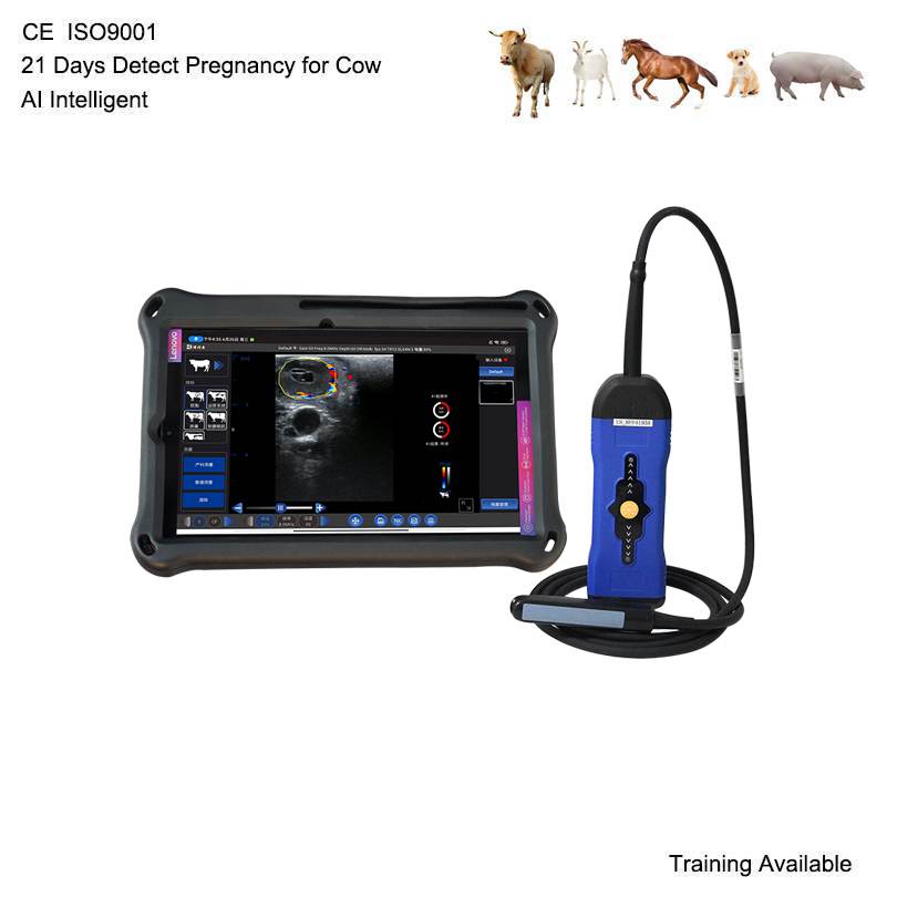
With a professional veterinary ultrasound scanner, cattle reproductive examinations can become more accurate, efficient, and informative.
Today, portable veterinary ultrasound machines, especially advanced wireless color Doppler ultrasound systems, are increasingly used in modern cattle reproduction programs.
1. What Is Cattle Reproductive Ultrasound?
Cattle reproductive ultrasound is a non-invasive imaging technique used to examine the reproductive organs of cows and heifers.
During a typical examination, a veterinarian inserts a suitable rectal ultrasound probe into the rectum to obtain images of the reproductive tract.
The ultrasound system converts reflected sound waves into real-time images, helping the operator evaluate different reproductive structures.
Common applications include:
– Uterine examination and reproductive tract evaluation.
– Ovarian ultrasound and follicle monitoring.
– Corpus luteum identification and assessment.
– Early pregnancy diagnosis in cattle.
– Embryo and gestational sac observation.
– Reproductive disorders and ovarian abnormalities.
– Monitoring follicular development during artificial insemination programs.
– Reproductive management in dairy and beef cattle.
For professional cattle reproduction, a suitable ultrasound scanner should provide clear imaging, reliable probe performance, convenient operation, and sufficient battery life for field examinations.
2. Ovarian Ultrasound: Monitoring Follicles and Reproductive Activity
Ovarian ultrasound is an important part of cattle reproductive management.
The ovaries contain follicles at different developmental stages. Monitoring their size, number, and development helps veterinarians understand ovarian activity and reproductive status.
Using a high-quality veterinary ultrasound machine, operators can examine:
Follicular development
Ultrasound imaging helps identify ovarian follicles and monitor their growth during the estrous cycle.
Dominant follicle monitoring
Identifying dominant follicles is useful when evaluating ovarian activity and planning reproductive management.
Corpus luteum examination
The corpus luteum (CL) develops after ovulation and plays an important role in progesterone production during the reproductive cycle.
Ovarian abnormalities
Ultrasound examination can assist in identifying ovarian cysts and other abnormalities that may affect reproductive performance.
For veterinarians involved in cattle breeding programs, clear ovarian images are particularly important for making informed reproductive management decisions.
3. Early Pregnancy Diagnosis in Cattle Using Ultrasound
Early pregnancy diagnosis is one of the most valuable applications of cattle reproductive ultrasound.
Traditionally, farmers and veterinarians have relied on rectal palpation and other diagnostic methods. Toutefois, ultrasound provides additional visual information about uterine contents and pregnancy development.
Depending on examination timing, equipment quality, probe selection, and operator experience, different pregnancy-related structures may become visible.
Here is a general overview:
Pregnancy stage Ultrasound examination
Around 21 days after AI Early pregnancy assessment may be possible with advanced equipment and experienced operators; confirmation can be challenging
Around 28–30 days Pregnancy-related fluid and gestational structures may become more identifiable
Around 35–45 days Embryo development and embryonic heartbeat can generally be evaluated
Around 45–60 days More developed embryonic structures can be observed
Important: These are approximate examination stages, not guaranteed detection windows. Pregnancy confirmation should follow appropriate veterinary examination protocols.