 BXL
Bovine Embryo Detection Ultrasound: Advanced Imaging for Early Pregnancy Diagnosis and Cattle Reproduction
1. Why Is Bovine Embryo Detection Ultrasound Important in Modern Cattle Breeding?
Efficient reproductive management is one of the most important factors influencing profitability in modern cattle farming. Whether managing a dairy herd, beef cattle operation, or professional breeding center, identifying pregnant and non-pregnant cows at the appropriate time helps farmers make better breeding and management decisions.
Traditional pregnancy diagnosis methods have limitations, particularly when veterinarians need to evaluate the reproductive tract and obtain more detailed information about early pregnancy.
This is where bovine embryo detection ultrasound becomes an important diagnostic technology.
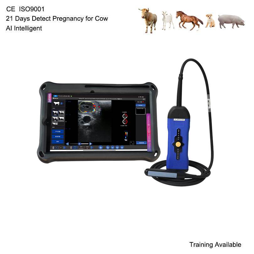
Veterinary ultrasound allows veterinarians to examine the uterus, Ovaires, and developing pregnancy structures in real time. Depending on the gestational stage, ultrasound imaging may help identify:
– Uterine fluid and changes associated with pregnancy.
– The gestational sac.
– The developing embryo.
– Embryonic heartbeat.
– Ovarian structures and corpus luteum.
– Changes in reproductive tissues.
Compared with methods that only determine whether an animal is pregnant, ultrasound provides additional anatomical information that supports more comprehensive reproductive examinations.
For veterinarians and cattle breeding professionals, a reliable bovine pregnancy ultrasound scanner is therefore a valuable tool for daily reproductive management.
2. How Does Bovine Embryo Detection Ultrasound Work?
Bovine embryo ultrasound uses high-frequency sound waves to produce real-time images of reproductive structures inside the cow.
A veterinary ultrasound probe transmits sound waves into the animal’s body. Different tissues reflect these waves differently, and the ultrasound system processes the returning signals to create images on the display.
For cattle reproductive examinations, transrectal ultrasound is widely used because it provides convenient access to the uterus and ovaries.
A suitable rectal linear probe can help veterinarians evaluate reproductive structures with detailed imaging.
During an examination, veterinarians may observe several important structures.
Gestational Sac
The gestational sac is an important early pregnancy structure that can be identified through ultrasound examination.
Its appearance and development provide useful information when evaluating early bovine pregnancy.
Embryonic Development
As pregnancy progresses, the embryo becomes visible within the gestational sac.
Ultrasound imaging helps veterinarians evaluate embryonic appearance and development according to gestational age.
Embryonic Heartbeat
At an appropriate stage of pregnancy, the embryonic heartbeat may be visualized using real-time ultrasound.
This provides additional information for pregnancy assessment and evaluation of embryonic viability.
Uterine Changes
The uterus undergoes structural changes during pregnancy.
Veterinarians can examine uterine contents, fluid distribution, and reproductive structures to support pregnancy diagnosis.
3. When Can You Detect Pregnancy and Embryos in Cattle Using Ultrasound?
One of the most common questions among cattle breeders is:
How early can you detect pregnancy in cows using ultrasound?
The detection time depends on ultrasound equipment, probe frequency, image quality, operator experience, examination technique, and the animal’s reproductive condition.
The following table provides a general reference for bovine reproductive ultrasound examinations.
Days after insemination Ultrasound examination
Around 21 days Early uterine changes may be evaluated, but routine pregnancy confirmation is not reliably established at this stage
25–28 days Early pregnancy structures may become detectable by experienced operators using suitable equipment
Around 28–30 days Gestational sac and embryo may be identifiable in suitable cases
30–35 days Pregnancy confirmation and embryonic assessment become more practical
35–45 days Embryonic structures and heartbeat can generally be evaluated more easily
After 45 days Further evaluation of embryonic development becomes possible
Important: These are approximate examination windows, not guaranteed detection dates. A negative scan at an early stage does not necessarily mean that the cow is not pregnant. Repeat examination may be necessary.
For commercial cattle farms, selecting an ultrasound scanner with stable image quality and a suitable rectal probe is essential for obtaining reliable diagnostic information.
4. What Features Should a Professional Bovine Embryo Ultrasound Scanner Have?
Not all veterinary ultrasound machines are equally suitable for cattle reproductive examinations.