Lameness in horses can severely affect performance, welfare, and productivity—whether the animal is a racehorse, a workhorse, or a breeding stallion. As a livestock farmer, I’ve experienced the frustration and concern that come when a horse shows signs of limping, uneven gait, or general discomfort. In my work, early diagnosis is everything. And for me and many others in the industry, ultrasound has become an essential tool in the battle against equine lameness.
Understanding Lameness in Horses
Lameness is any alteration in a horse’s gait, usually caused by pain or mechanical dysfunction. It can stem from a wide variety of sources—tendon injuries, ligament damage, Problemas articulares, or even abscesses in the hooves. While traditional clinical examination, hoof testers, and nerve blocks are useful, they don’t always give the complete picture. That’s where diagnostic imaging comes into play.
Why Ultrasound?
Ultrasound imaging—or ultrasonography—has become one of the most commonly used diagnostic tools in veterinary medicine. It uses high-frequency sound waves, typically between 1.5 y 18 megahertz (MHz), to visualize internal structures. These sound waves are emitted from a transducer, pass through the body, and are reflected back by different tissues. The resulting echoes are converted into detailed real-time images.
Compared to radiography (X-rays), which is better at showing bones, ultrasound excels at evaluating soft tissue structures. That makes it ideal for identifying tendon tears, ligament injuries, joint capsule swelling, and other subtle musculoskeletal abnormalities that often underlie lameness.
My Personal Experience Using Ultrasound for Equine Lameness
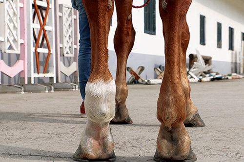
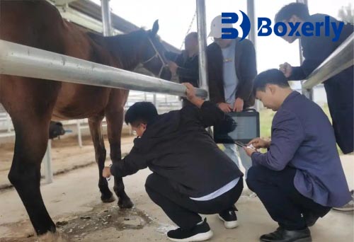
When one of my mares started showing mild lameness in her left forelimb, I initially suspected it was just a minor strain. There was no swelling, no heat, and the lameness was intermittent. Sin embargo, based on past experience, I knew better than to ignore the early signs. I scheduled an ultrasound examination.
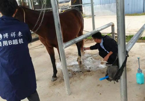
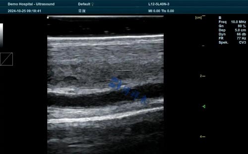
The vet used a high-frequency linear transducer and applied coupling gel to ensure clear transmission of sound waves. As the probe moved down the leg, real-time B-mode images appeared on the screen. These grayscale images clearly showed the digital flexor tendons, suspensory ligament, and surrounding tissues.
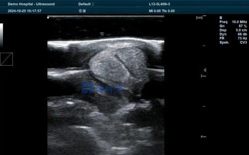
To my surprise, there was a partial lesion in the superficial digital flexor tendon—something not visible externally and undetectable via palpation. Thanks to early detection, we implemented rest, controlled exercise, and physiotherapy. That mare made a full recovery and went on to have a successful foaling season.
Ultrasound in Practice: What It Can Show
In horses, lameness often originates from injuries to the following areas:
-
Flexor tendons (superficial and deep digital)
-
Suspensory ligaments
-
Check ligaments
-
Annular ligaments
-
Joint capsules
-
Bursa and synovial sheaths
Ultrasound allows for precise visualization of these structures. Not only can it detect lesions and fluid accumulation, but it also allows repeated monitoring during treatment, helping track healing progress over time.
Because ultrasound works in real time, I can observe changes immediately, and we can make quicker decisions. Plus, it’s non-invasive, safe, and can be done stall-side with minimal stress for the animal.
Equipment Considerations for Equine Ultrasound
Not all ultrasound machines are the same. For musculoskeletal imaging, a linear-array transducer with frequencies between 7.5–15 MHz is commonly used. This provides high-resolution images of superficial structures—perfect for tendons and ligaments.
Sometimes, a lower frequency probe (like 3.5–5 MHz) may be used to image deeper structures, but with lower image resolution. In my experience, choosing the right transducer is crucial. I always make sure my vet uses the right probe for the target depth and adjusts the gain, centro de atención, and depth settings accordingly.
Benefits of Early Diagnosis
The earlier a lesion is identified, the better the chance of full recovery. Ultrasound has helped me prevent minor tendon strains from turning into career-ending injuries. It’s also helped guide treatment—whether it’s rest, anti-inflammatory medications, shockwave therapy, or regenerative techniques like stem cell injections.
For breeding stallions and broodmares, ultrasound is invaluable. Even subtle injuries can affect performance or lead to complications during breeding. By using ultrasound, I’ve been able to keep my animals sound, reduce downtime, and plan treatments with more precision.
Advancing Uses: Guided Interventions
Ultrasound isn’t just for diagnosis. In skilled hands, it’s also a tool for guiding therapeutic procedures:
-
Ultrasound-guided injections (like corticosteroids or regenerative therapies) into tendon sheaths or joints.
-
Fine-needle aspiration or biopsies to diagnose infections or tumors.
-
Monitoring healing progression through regular follow-up scans.
These guided techniques reduce the risk of complications and improve the accuracy of treatment delivery.
Limitations and Challenges
As powerful as ultrasound is, it does have its limitations. Bone, for example, reflects most of the ultrasound waves, so we can only see the surface—not the interior. That’s why for suspected fractures or deep joint issues, radiographs or CT might be necessary.
Adicionalmente, interpreting an Imagen de ultrasonido requires skill and experience. An inexperienced operator might miss a subtle lesion or mistake a normal variation for pathology. That’s why I always work with a vet who’s trained in equine ultrasonography.
Integrating Ultrasound into Daily Farm Management
Hoy, I consider ultrasound a key part of my farm’s veterinary strategy. Whether for routine reproductive checks, lameness evaluation, or monitoring a healing injury, it’s an invaluable tool that helps reduce guesswork and improve outcomes.
Over the years, I’ve become familiar with basic image interpretation—recognizing tendon fiber alignment, fluid pockets, or irregular margins. This has helped me communicate more effectively with veterinarians and better understand the condition of my animals.
Final Thoughts
Ultrasound has revolutionized how we detect and manage lameness in horses. From pinpointing invisible soft tissue injuries to guiding targeted therapies, it’s become an essential part of maintaining equine health and performance. For farmers and breeders like me, it offers peace of mind and helps protect the animals we depend on.
In my experience, the investment in ultrasound diagnostics pays for itself many times over—in healthier animals, faster recoveries, and fewer long-term complications.