Hinchazón en perros, particularmente en caninos de trabajo o de granja, no solo es incómodo, sino que puede poner en peligro la vida. A medida que crece la demanda mundial de diagnósticos veterinarios, especialmente en entornos rurales y agrícolas, ultrasound imaging has become a valuable tool for evaluating abdominal conditions in dogs. But can a dog receive an ultrasound specifically for bloating? Absolutely—and in many cases, they should.

En este artículo, we will explore the relationship between canine bloating and veterinary ultrasound, the science behind why and how ultrasounds are used, the procedures involved, and what farm and livestock professionals should know to ensure their dogs receive timely, effective care.
Understanding Bloating in Dogs
What is Canine Bloating?
Canine bloating—commonly referred to as Gastric Dilatation and Volvulus (GDV)—is a condition where a dog’s stomach fills with gas, fluid, or food and may then twist. This twisting (volvulus) can cut off blood supply to the stomach and surrounding organs, causing severe pain, tissue death, and potentially death if not treated immediately.
Though GDV is most commonly seen in deep-chested breeds like Great Danes, German Shepherds, and Dobermans, it can occur in any dog. For working dogs or herding breeds commonly used on farms, early detection is vital.
Signs and Symptoms
Typical signs of bloating include:
-
A visibly distended abdomen
-
Unsuccessful attempts to vomit or belch
-
Restlessness or pacing
-
Excessive drooling
-
Labored breathing
-
Weakness or collapse
These symptoms can progress rapidly. Immediate veterinary attention is critical, and ultrasound is often part of the diagnostic approach.
How Ultrasound Helps Diagnose Bloating in Dogs
The Role of Veterinary Ultrasound
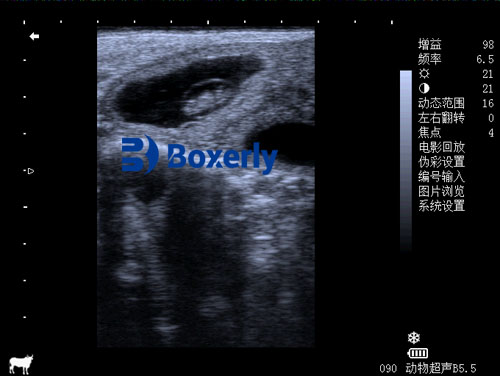
Ultrasound uses high-frequency sound waves to produce real-time images of internal structures. In cases of bloating, ultrasound provides valuable insights into:
-
The degree of gastric distension
-
Whether volvulus (stomach twisting) has occurred
-
Compromised blood flow to organs
-
The presence of fluid, gas, or foreign bodies
-
Damage to spleen or other abdominal structures
Unlike X-rays, which give static, less-detailed images, ultrasound offers dynamic, real-time observation—making it ideal for rapidly assessing critical abdominal conditions like GDV.
When Is Ultrasound Used?
Veterinarians may recommend an abdominal ultrasound in any of the following cases:
-
The dog has persistent or worsening abdominal bloating
-
There are inconclusive results from physical exams or X-rays
-
The clinical team needs to determine whether surgery is necessary
-
To monitor progress post-surgery or during recovery
Advantages of Ultrasound in Diagnosing Bloating
Veterinary ultrasound presents several key advantages over other diagnostic tools, particularly in field or farm settings:
-
Non-invasive: No incisions or anesthesia are required in most cases.
-
Real-time imaging: Allows clinicians to observe organ movement, flujo sanguíneo, and gas patterns instantly.
-
Portable technology: Modern ultrasound units are lightweight and battery-operated, making them ideal for mobile veterinary practice.
-
Safe and repeatable: Can be used multiple times without radiation exposure.
For farm operators or handlers of working dogs, the accessibility and safety of ultrasound make it an invaluable diagnostic tool.
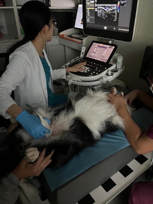
How the Ultrasound Procedure Works in Dogs
Preparing the Dog
If bloating is suspected, time is critical. Most veterinarians prioritize stabilizing the dog—using IV fluids, decompressing the stomach, and administering pain relief—before conducting an ultrasound. Sin embargo, when the dog is stable, the procedure typically involves:
-
Ayuno: Not necessary in emergencies but preferred if time allows.
-
Shaving the abdomen: Hair is shaved to allow better contact with the probe.
-
Application of gel: A conductive gel is applied to eliminate air pockets between the probe and the skin.
Performing the Ultrasound
Using a micro-convex or linear probe (3.5–8 MHz), the veterinarian scans the abdominal cavity. Areas of focus typically include:
-
Stomach: Checking for dilation, abnormal position, and wall thickness.
-
Spleen and liver: Assessing for torsion or blood supply compromise.
-
Intestines: Looking for obstructions or abnormal motility.
-
Free fluid: Identifying peritoneal effusion or hemorrhage.
Images are interpreted in real time. If torsion is confirmed, emergency surgery may be initiated immediately.
Case Example: Ultrasound in Rural Practice
Consider a scenario on a livestock farm in Argentina, where a working Border Collie exhibits signs of bloating after herding cattle in extreme heat. The nearest urban veterinary hospital is over 100 kilometers away. Sin embargo, the local veterinary technician is equipped with a portable ultrasound unit.
Within minutes, the technician confirms gastric dilatation without volvulus. The dog is stabilized on-site, and preventive measures are taken—potentially saving the animal’s life and preserving a valuable working partner.
This scenario underscores the importance of Ecografía veterinaria in field-based animal healthcare.
Preventive Use of Ultrasound in Bloat-Prone Dogs
While ultrasound is invaluable in emergencies, it can also serve a preventive role:
-
Monitoring gastric motility: In dogs with recurring GI issues.
-
Screening before breeding: Especially for breeds prone to bloat.
-
Post-surgical checks: After gastropexy or other abdominal surgeries.
-
Dietary impact assessments: Checking digestion patterns in performance dogs.
Integrating routine ultrasound exams into canine health protocols can support long-term wellness and reduce the risk of acute bloating episodes.
Choosing the Right Ultrasound Machine for Canine Diagnostics
For veterinarians or farm operators looking to invest in ultrasound equipment, especially for canine and small animal use, consider the following:
1. Probe Type
-
Micro-convex probes: Excellent for small to medium-sized dogs due to their smaller footprint.
-
Linear probes: Ideal for superficial structures and high-resolution imaging.
2. Frequency Range
-
A range of 5–8 MHz is suitable for most canine abdominal applications.
3. Portabilidad
-
Choose battery-operated or handheld systems for farm or mobile practice.
4. Modos de imagen
-
B-mode: Standard grayscale imaging.
-
Color Doppler: Useful to assess blood flow, particularly in torsion cases.
-
M-mode and 3D/4D imaging (optional): For advanced diagnostic capabilities.
5. Durability and Ease of Use
-
Equipment designed for rugged, field conditions (like BXL’s veterinary series) is ideal for rural use.
Veterinary and Ethical Considerations
Bloating in dogs is painful and often fatal if left untreated. Timely diagnostics are not just a matter of convenience—they’re a matter of animal welfare. Farm operators have both a practical and ethical responsibility to ensure that their animals, including dogs, receive the best possible care.
Ultrasound allows early detection, accurate diagnosis, and faster intervention, all of which contribute to improved animal outcomes and lower long-term costs.
Final Thoughts
Sí, dogs can and should receive an ultrasound when bloating is suspected. This technology, long trusted in human medicine and large animal veterinary practice, is equally indispensable for diagnosing abdominal emergencies in canines.
For those in livestock and rural animal operations, investing in or partnering with veterinary professionals who utilize ultrasound technology is not a luxury—it’s a modern necessity. Whether for emergency diagnosis, routine monitoring, or post-surgical care, ultrasound stands as one of the most valuable tools in canine health management today.