As a modern dairy farmer or veterinarian, understanding the dynamics of follicular development in dairy cows is essential for optimizing reproductive efficiency. One of the most effective technologies for achieving this is ultrasound imaging—a non-invasive, real-time method that allows for precise evaluation of ovarian follicles at different stages. Compared to traditional rectal palpation, ultrasound offers significantly improved accuracy, especially for small follicles and complex ovarian structures.
في هذه المقالة, we explore how ultrasound is used to monitor follicular development in dairy cows, the insights it provides into ovarian physiology, and how this tool supports advanced reproductive management across various farming systems.
Why Ultrasound Is the Preferred Method
Ultrasound imaging has revolutionized the way reproductive health is monitored in dairy herds. In particular, transrectal ultrasonography allows veterinarians to detect follicles as small as 2–3 mm in diameter—far below the detection threshold of manual rectal palpation. Research shows that the detection rate of follicles larger than 10 mm reaches up to 92% using ultrasound, compared to only 62% using rectal examination (Ginther et al., 1995).
Even more impressively, for follicles larger than 15 المليمتر, ال detection rate via ultrasound reaches 100%. These levels of precision are crucial when managing synchronization protocols or assessing follicular dynamics in cows undergoing hormonal treatments such as superovulation.
In field settings across North America and Europe, ultrasound has become the standard for monitoring ovarian activity, due to its ability to provide live morphological images of follicular waves, corpus luteum development, and ovulation timing.

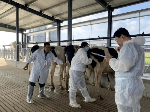
Ultrasound imaging of cow ovarian follicles
Understanding Follicular Waves in Dairy Cows
The estrous cycle of dairy cows typically consists of 2–4 follicular waves. A large-scale study conducted in Canada found that approximately 71.4% of cows have three follicular waves per cycle, while 21.3% experience four (Adams et al., 1992). Each wave generally lasts 7 ل 9 أيام, shorter than the 13-day persistence of cystic follicles.
During each wave, a cohort of small follicles emerges, and one follicle is selected to become dominant. With ultrasound, these phases can be closely tracked. For instance, in early antral stages, follicles range from 1 ل 4 mm in diameter. As they grow, mid-size follicles reach 5–7 mm, and dominant follicles exceed 8 المليمتر. At ovulation, the preovulatory follicle typically reaches a diameter of 14–22 mm.
In one study involving 20 cyclic Holstein cows, ultrasound monitoring showed that the largest dominant follicle had an average diameter of 13.6 المليمتر, while mature ovulatory follicles reached 13.8 mm on average. These precise measurements help determine the cow’s readiness for breeding or artificial insemination.
Dynamic Follicular Development in Young Cows
Ultrasound also plays a key role in monitoring follicular development in heifers and young cows. A longitudinal study found that young dairy cows exhibited high follicular activity, with an average of 51.66 small follicles per cycle, making up more than 92% of the ovarian follicular population.
The continuous tracking of follicular growth in these cows revealed dynamic changes across the estrous cycle. For instance, on day 2 of the cycle, dominant follicles averaged 8.3 mm in diameter. By day 5, they grew to 13.5 المليمتر. In cows undergoing superovulation treatments, growth was even more dramatic, with follicle size increasing from 14.5 mm on day 10 ل 20.3 mm on day 12.
Interestingly, the thickness of the follicular wall decreased from 4 mm to 2 mm during this period—a key sign of imminent ovulation. These changes can be accurately detected only through high-resolution ultrasound.
Follicular Morphology Across Estrous Stages
Ultrasound allows researchers and practitioners to visualize the changes in follicle size and morphology throughout the estrous cycle. Dominant follicles average 11.1 mm in the selection phase, expand to 14.2–15.9 mm in the estrus phase, and reach 21 mm just before ovulation. When this threshold is reached, cows typically enter the ovulatory stage.
One study conducted in Europe examined the left and right ovaries separately. It found that the average diameter of the largest dominant follicle was 1.27 ± 0.29 cm on the left ovary and 1.32 ± 0.23 cm on the right ovary. Furthermore, cows had approximately 2.60 dominant follicles on the left and 2.47 on the right—data that would be impossible to obtain using manual palpation alone.
Integrating Hormone Profiling with Ultrasound
While ultrasound provides detailed morphological information, combining it with blood hormone profiling offers a more complete understanding of follicular dynamics. Using automated, remote-controlled blood sampling devices, farmers can collect high-frequency blood samples for progesterone and estrogen analysis.
These hormonal levels, correlated with ultrasound data, help determine the precise timing of follicle selection, dominance, and ovulation. In regions such as the Netherlands, ألمانيا, and Japan, this integrated approach is increasingly used to improve fertility rates through timed artificial insemination (TAI) و estrus synchronization programs.
Moreover, the combined strategy helps in studying breed-specific follicular patterns and how environmental conditions—such as heat stress or photoperiod changes—affect reproductive performance.
Benefits of Ultrasound-Based Follicular Monitoring
1. الكشف المبكر عن التشوهات
Ultrasound can identify cystic follicles, luteal insufficiency, and anovulatory cycles early. This enables timely veterinary intervention and improves conception rates.
2. Accurate Ovulation Prediction
Follicle diameter is a reliable predictor of ovulation. By tracking follicle size and wall thickness, farmers can precisely schedule insemination for optimal fertility.
3. Enhanced Superovulation Management
During superovulation programs, ultrasound is essential to track follicle recruitment and avoid overstimulation or follicular cysts.
4. Cost-Efficient Breeding Decisions
Timely identification of dominant follicles reduces unnecessary inseminations, cuts hormonal treatment costs, and increases pregnancy rates.
5. Improved Genetic Selection
By monitoring ovarian response to synchronization protocols, ultrasound helps identify cows with superior reproductive performance—crucial for genomic selection.
Future Trends and Global Adoption
Globally, the adoption of ultrasound in dairy farming is increasing. In North America, over 80% of large dairy operations now use ultrasound for reproductive monitoring. In Europe, portable ultrasound devices are routinely used by veterinarians visiting farms.
Meanwhile, in developing regions such as Latin America and Southeast Asia, low-cost ultrasound units are enabling smallholder farmers to access high-quality reproductive diagnostics. As devices become more affordable and training more widespread, ultrasound will likely become a universal standard in herd fertility management.
استنتاج
Ultrasound monitoring of follicular development in dairy cows has become an indispensable tool for modern herd management. With its unmatched precision, التصوير في الوقت الحقيقي, and ability to detect subtle changes in follicular dynamics, ultrasound empowers farmers and veterinarians to make informed, efficient, and data-driven breeding decisions.
Whether tracking follicular waves, planning insemination, or managing synchronization protocols, ultrasound is setting a new standard in reproductive efficiency. As research deepens and technology becomes more accessible, its role in shaping the future of dairy fertility continues to expand—bringing productivity, profitability, and animal welfare to new heights.
References
-
Ginther, O. J., & Kot, K. (1995). Follicular dynamics during the bovine estrous cycle. Theriogenology, 43(6), 1211-1226.
https://doi.org/10.1016/0093-691X(95)00080-E -
Adams, G. P., Matteri, R. L., Kastelic, J. P., Ko, J. C. H., & Ginther, O. J. (1992). Association between surges of follicle-stimulating hormone and the emergence of follicular waves in heifers. Journal of Reproduction and Fertility, 94(1), 177–188.
https://doi.org/10.1530/jrf.0.0940177 -
Smith, E. R., & Whitaker, D. A. (2021). Veterinary Ultrasonography in Food-Producing Animals. Journal of Veterinary Imaging.
https://www.jvimaging.org/article/ultrasound-cattle-follicles