Imagine chatting with a friendly rancher out in the field, swapping stories over coffee—this is how I’m approaching this topic. Talking about using ultrasound on beef cattle isn’t stiff; it’s practical, full of real-life value, and down-to-earth.
Why ultrasound matters on modern beef farms
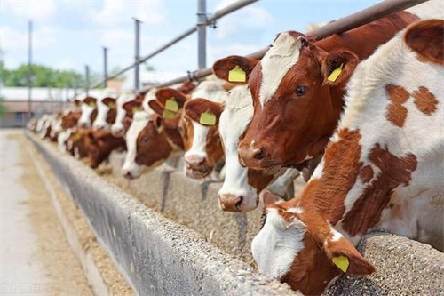
As a livestock farmer, I’ve learned that knowing exactly where each animal is in its growth journey makes decisions a whole lot easier. Western producers in the UGA Cooperative Extension, and researchers in Lithuania and Canada, all point in the same direction—ultrasound helps monitor muscle area, Шпик, rump fat, and even intramuscular fat (marbling) while cattle are still alive.
Farmers in the US have used this handy system for decades. It made bull-progeny testing take just two years instead of seven and cut costs from around $5,000 Кому $1,000 per sire. In Lithuania, a study on Black Angus found that bulls hitting about 430 kg already showed high marbling via ultrasonography—and around 600 kg for heifers.
Growth phases and what ultrasound reveals
Young beef cattle go through that familiar S‑shaped growth curve. Fast gain early on, then muscle increases slow as fat starts piling on. The longissimus dorsi muscle (the ribeye) is key. Measuring eye muscle area (or ribeye area) at the 12th–13th rib shows muscle growth trends nicely. Fat thickness at the rib or rump also indicates readiness for finishing.
Breed matters. Angus, which mature early, tend to show plateau in muscle sooner, while Charolais or Simmental (later‑maturing breeds) keep building muscle longer.
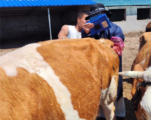
How I run scans on the farm
-
Use a real‑time linear probe (2–10 MHz), with a gel to get clear images.
-
Point it between the 12th and 13th ribs to capture the longissimus dorsi area.
-
Measure backfat depth and rump fat, then estimate intramuscular fat.
-
If you do scans serially (e.g. every month), you see trends—when muscle slows, when fat picks up, и так далее.
Here’s a basic representation of what data I pull:
| Parameter | What it tells me | Where measured |
|---|---|---|
| Ribeye area (cm²) | Muscle growth potential | Between 12th and 13th ribs |
| Backfat thickness (миллиметр) | External fat, finish level | Rib region |
| Rump fat (миллиметр) | Extra cover & finish | Between hooks and pins |
| Estimated marbling (%) | Intramuscular fat & meat quality | Same scan area, visual estimate |
Real‑world benefits farmers get
-
Non‑invasive and safe
No blood, no biopsy, just sound waves. Animals aren’t stressed. -
Quantitative insight
Gives exact measurements for comparing individuals, making culling or finishing easier. -
Real‑time decisions
When you scan and see that ribeye area is plateauing but fat’s climbing, you know it’s time to switch to finishing or market weight. -
Breeding and genetics
By scanning young bulls or replacements, you can select animals with better carcass trait potential earlier.
International perspective: how western researchers see it
-
In the US, Extension publications emphasize ultrasound as an economical tool to improve carcass genetics fast, replacing slow traditional methods.
-
Canadian research with young bulls across Angus, Charolais, Hereford, and Simmental showed consistent breed differences in muscle and fat via ultrasound scans reliablly predicting end performance.
-
Lithuanian studies in 2025 confirmed scans can monitor marbling progression through weight ranges and support selecting for meat quality traits before slaughter age.
Matching feeding strategy to growth stage
When a calf is in the youth phase—post-puberty, but not mature—it’s muscle time. I adjust protein-rich feeding to support lean growth. Once the ribeye area levels off, I shift to energy-dense finishing rations to boost marbling and external fat. This tailored feeding strategy avoids over‑feeding or wasting feed.
Breed plays in again: early-maturing breeds peak muscle earlier, later ones benefit from extended lean feeding.
Reproduction uses too (they go beyond growth)
While most of us focus on scanning for growth, vets and reproductive managers use ultrasound for pregnancy checks too:
-
B‑mode scans help confirm pregnancy early, assess uterine structure.
-
Doppler mode shows blood flow to corpus luteum and follicles, which helps detect pregnancy or evaluate embryo‑recipient readiness.
-
Color Doppler helps with early pregnancy diagnosis around day 20, enabling faster resynchronization in breeding programs.
Though that’s more for reproductive herds than feedlots, it’s a good illustration of how versatile ultrasound tech is.
A little chatty wrap-up
Honestly, I’ve found few tools more practical than ultrasound. It’s like a peek inside each animal without doing harm. Measurement data means smarter, faster decisions. Feed to the right stage. Cull or finish at the right time. And for breeding herds, ultrasound supports reproductive efficiency too.
More and more producers outside North America are adopting it too. The blend of real-time data, non-invasive scanning, and genetic evaluation makes it too good to pass up.