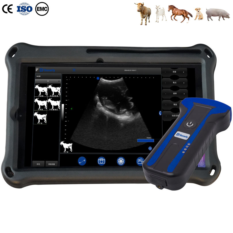
Instructions for Using a Swine Ultrasound Machine to Detect Backfat Thickness, Eye Muscle Area, and Intramuscular Fat
Preparation for Using a Swine Ultrasound Machine: When using a swine ultrasound machine to detect backfat thickness, place the ultrasound probe horizontally on the pig’s back using a water-resistant bag. The probe axis should be perpendicular to the midline of the back. The scanning line of the ultrasound probe should slightly extend beyond the midline of the back to visualize the strong echoes of the spinous processes. To measure the eye muscle area, use a swine ultrasound probe with a scanning width of at least 10cm. A water-resistant bag should be placed between the ultrasound probe and the pig’s back. Adjust the contrast, brightness, прибыль, and near-field/far-field settings of the ultrasound machine to obtain a clear ultrasound image. Freeze the image and use the electronic distance and area measurement functions to measure the backfat thickness and eye muscle area. Add annotations such as ear tag, breed, age, and weight using the annotation function. Finally, print or input the data into a computer for storage.
Poor quality images acquired using a swine ultrasound machine may exhibit the following characteristics: ① Blurred images, caused by the pig moving during image freezing or the technician moving the sensor. ② Poor contact between the sensor and skin, possibly due to pig movement during image freezing or insufficient application of coupling agent or vegetable oil. ③ Dark areas in the ultrasound image, caused by uneven pressure applied to the sensor by the technician. ④ Bright echoes or periodic repetitive lines of electrical interference, caused by strong interfacial reflections between fat layers, which can be minimized by slightly tilting the sensor over the skin surface.
A clear swine ultrasound image clearly shows four strong echo points: the spinous process, vertebral body, transverse process, and longitudinal fascia of the lateral end of the longissimus dorsi muscle, as well as four strong echo bands: the skin interface, interfacial connective tissue, and the upper and lower fascia of the longissimus dorsi muscle. The fat layer appears as a band-shaped, solid, dark area, while the muscle section appears as a slightly more echogenic, solid, dark area. The measurement point for backfat thickness and eye muscle thickness is 4 cm from the spinous process. The distance between the skin interface and the two strong echogenic bands of the upper muscle membrane is the backfat thickness. On a pig ultrasound machine, the distance between the strong echogenic bands of the upper and lower muscle membranes is the solid dark area surrounded by the four strong echogenic points on both sides and the strong echogenic bands of the upper and lower muscle membranes, as measured by the muscle thickness and eye muscle area measurement area.
Backfat thickness and eye muscle area are directly related to lean meat percentage. They are highly valued as two important indicators in pig genetic breeding and performance evaluation, and their accurate measurement is of great significance. To ensure the accuracy and stability of measuring backfat thickness and eye muscle area in live pigs using a pig ultrasound machine, the personnel must practice repeatedly and patiently, comparing live and carcass samples, to achieve proficiency and accuracy. The correlation between the results measured by two different personnel must be at least 95% to prevent significant errors in measurement scores due to subjective differences when taking turns measuring. For backfat thickness and eyelid muscle area at the same location on the same individual, at least three sets of data must be analyzed, and the coefficient of variation of the three analyses must not exceed 5%. Otherwise, data with excessively large discrepancies will be considered invalid.