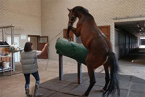
Artificial insemination (AI) has revolutionized livestock reproduction worldwide, providing farmers with efficient, cost-effective methods to improve genetics, control breeding schedules, and increase herd productivity. しかし, successful AI relies heavily on precise ovulation timing, which can be challenging due to the natural variability in animals’ estrous cycles. To address this, veterinarians and reproductive specialists use various ovulation induction protocols to synchronize estrus and ovulation, optimizing conception rates.

In this context, veterinary ultrasound technology has emerged as a vital tool to monitor ovarian dynamics, track follicular growth, and accurately evaluate the efficacy of different ovulation induction protocols. This article explores how veterinary ultrasound tech is used to compare ovulation induction protocols in artificial insemination, emphasizing practical applications, scientific insights, and global perspectives.
Understanding Ovulation Induction Protocols in Artificial Insemination
The Challenge of Estrus Synchronization
Natural estrus cycles in livestock, particularly cattle, 羊, and goats, vary widely between individuals and breeds. This variability can lead to missed breeding windows, inefficient insemination, and suboptimal pregnancy rates. Synchronization protocols are therefore developed to control or induce ovulation at predictable times, allowing timed artificial insemination (TAI) without the need for estrus detection.
Common Ovulation Induction Protocols
Several ovulation induction protocols are widely used across the globe, with regional preferences shaped by breed characteristics, farm management, and available technologies. The main protocols include:
-
Prostaglandin (PGF2α)-based Protocols: These induce luteolysis to regress the corpus luteum and trigger a new estrous cycle.
-
GnRH-based Protocols: Gonadotropin-releasing hormone is used to induce follicle ovulation or luteinization.
-
Ovsynch Protocol: Combines GnRH and PGF2α injections to synchronize follicular waves and ovulation precisely.
-
CIDR (Controlled Internal Drug Release) with Hormonal Treatments: Uses progesterone releasing devices combined with PGF2α and/or GnRH to control the cycle tightly.
Each protocol varies in complexity, duration, 費用, and efficacy, and choosing the right one depends on species, breed, and farm goals.
The Role of Veterinary Ultrasound in Comparing Ovulation Induction Protocols
Ultrasound as a Monitoring Tool
Veterinary ultrasound offers a non-invasive, real-time visualization of ovarian structures—follicles, corpora lutea, and cysts—providing critical information on follicular growth dynamics and luteal function. This ability to monitor internal reproductive organs directly has transformed how veterinarians assess the physiological response to ovulation induction.
実際に, ultrasound enables:
-
Follicular Size Measurement: Determining the dominant follicle size to predict ovulation readiness.
-
Detection of Ovulation: Confirming rupture of follicles and formation of corpus luteum.
-
Luteal Function Assessment: Evaluating corpus luteum development and progesterone production indirectly.
These observations help to compare the effectiveness of different protocols in synchronizing ovulation and improving AI timing.
Case Studies in Protocol Comparison Using Ultrasound
-
Comparing Ovsynch with PGF2α Protocols in Dairy Cows
A study conducted in the US utilized ultrasound to monitor follicular development in Holstein dairy cows undergoing either Ovsynch or PGF2α protocols. Ultrasound scans revealed that cows in the Ovsynch group had more synchronized follicular wave emergence and ovulation, leading to higher pregnancy rates compared to those receiving PGF2α alone.
-
GnRH versus CIDR-Based Protocols in Beef Cattle
Research from Australia showed that ultrasound monitoring of Angus and Hereford cows undergoing GnRH or CIDR-based protocols demonstrated more consistent follicle maturation and luteal formation in CIDR-treated cows, resulting in more predictable ovulation times and improved AI success.

How Ultrasound Enhances Protocol Optimization
Real-Time Feedback on Hormonal Treatments
Ultrasound provides direct feedback on how animals respond to hormonal injections. 例えば, if follicles fail to develop or ovulate as expected, veterinarians can adjust dosages, timing, or switch protocols, maximizing conception rates.
Early Identification of Non-Responders
Some animals do not respond optimally to induction protocols due to underlying reproductive disorders such as cystic ovaries or silent heat. Ultrasound allows early detection of these issues, enabling targeted veterinary interventions or removal from breeding programs, reducing wasted inseminations.
Economic and Welfare Benefits
By improving synchronization accuracy and conception rates, ultrasound-guided protocol selection reduces the number of inseminations per pregnancy, cutting semen and labor costs. It also minimizes animal stress by avoiding repeated handling and hormonal treatments.
Global Perspectives and Innovations in Veterinary Ultrasound Tech
Adoption in Developed versus Developing Regions
In technologically advanced countries such as the United States, カナダ, オーストラリア, and parts of Europe, veterinary ultrasound machines are standard tools on commercial farms and veterinary practices. Integration with precision livestock farming systems enables automated 生殖モニタリング, improving AI outcomes further.
In developing regions, ポータブル, affordable ultrasound devices have increased access to reproductive management tools, helping smallholder farmers optimize breeding despite limited resources.
Advances in Ultrasound Imaging and AI Integration
Recent technological advancements include:
-
3D and Doppler Ultrasound: Offering enhanced visualization of blood flow in ovarian tissues, providing insights into follicle health and luteal function.
-
Artificial Intelligence (AI) Algorithms: Automating follicle detection and ovulation prediction, reducing operator dependence.
These innovations promise to further refine ovulation induction protocol comparisons, making data-driven decisions faster and more reliable.
Practical Recommendations for Farmers and Veterinarians
Selecting an Ovulation Induction Protocol
Consider species, breed, farm size, and available labor. For high-value dairy herds, Ovsynch combined with ultrasound monitoring may deliver best results. For extensive beef operations, simpler PGF2α protocols might suffice but benefit from ultrasound confirmation.
Incorporating Ultrasound into Routine Reproductive Management
Train veterinary staff or farmers in basic ultrasound techniques to monitor follicular development regularly. Implement scans at critical points during the protocol, such as before AI and 7–10 days post-AI to confirm ovulation and early pregnancy.
Data-Driven Continuous Improvement
Record ultrasound findings alongside insemination and pregnancy results. Analyze this data to refine protocol choice and timing annually, adapting to herd-specific reproductive patterns.
結論
Veterinary ultrasound technology has revolutionized the way ovulation induction protocols are evaluated and applied in artificial insemination programs. By offering direct, non-invasive insights into ovarian function, ultrasound allows practitioners to compare protocols effectively, tailor reproductive management, and improve conception rates.
Globally, both in advanced and emerging livestock systems, ultrasound-guided synchronization protocols are improving animal welfare, economic efficiency, and genetic progress. As ultrasound devices become more portable, 手頃 な 価格, and technologically sophisticated, their integration with AI and digital livestock management platforms will further enhance reproductive success in the future.
For livestock farmers and veterinarians aiming to maximize the success of artificial insemination, investing in veterinary ultrasound technology and understanding protocol differences is not just a scientific advantage—it’s a practical necessity.
References
-
Pursley JR, Mee MO, Wiltbank MC. Synchronization of ovulation in dairy cows using PGF2α and GnRH. Theriogenology. 1995 Jul;44(7):915-23. DOI: 10.1016/0093-691X(95)00206-7
URL: https://doi.org/10.1016/0093-691X(95)00206-7 -
Carvalho PD, et al. Use of ultrasonography to evaluate the Ovsynch protocol in beef cattle. Animal Reproduction Science. 2012;132(1-2):34-40.
URL: https://doi.org/10.1016/j.anireprosci.2012.01.005 -
Stevenson JS, et al. The impact of synchronization protocols on follicular dynamics and fertility in dairy cows. Journal of Dairy Science. 2004;87(10):2887-95.
URL: https://doi.org/10.3168/jds.S0022-0302(04)73475-4 -
Sterry RA, et al. Advances in reproductive ultrasonography in cattle. Veterinary Clinics: Food Animal Practice. 2010;26(1):63-76.
URL: https://doi.org/10.1016/j.cvfa.2009.11.004 -
Baruselli PS, et al. Synchronization and timed artificial insemination in beef cattle: practical applications and protocols. Theriogenology. 2017;104:1-10.
URL: https://doi.org/10.1016/j.theriogenology.2017.07.001