Correctly recognizing the images of veterinary B-ultrasound machines to avoid misjudgments during the inspection process is a vital technology for breeding farms, veterinary stations, service units and scientific research units. The images of veterinary B-ultrasound machines used are not clear, or It is the misjudgment of the B-ultrasound image, which will directly affect the daily management efficiency and production efficiency. There are many reasons for the misjudgment caused by the operation of the veterinary B-ultrasound machine. The experts of Boxiang come to the veterinary B-ultrasound machine to explain to you the misjudgment various reasons.
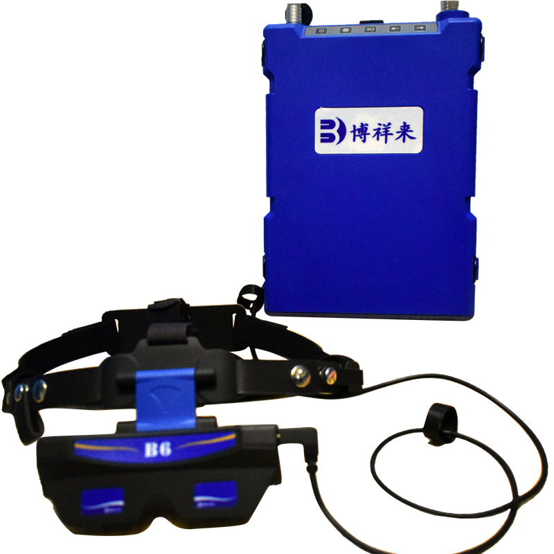
Bo Xianglai’s latest veterinary ultrasound machine BXL-V60 for cows and sheep .

1 .Determined by the performance and resolution of the veterinary ultrasound machine itself
Choosing a good B-ultrasound can get very clear images during the follicular development check to grasp the mating timing and the pregnancy check, which will greatly improve the accuracy and advance the check time. When many users buy veterinary B-ultrasounds, they think about the cheapness, but they don’t know that the performance of cheap B-ultrasounds may affect the diagnosis results, so I also suggest users and friends not to make them cheap for a while. , Be sure to buy a veterinary B-ultrasound machine suitable for you according to your own needs according to the tested animals and the desired purpose.
2.The operator has insufficient experience in using the veterinary ultrasound machine or does not understand the imaging principle of the ultrasound machine at all
The veterinary B-ultrasound machine is a high-tech B-ultrasound diagnostic equipment. It requires proficient experience to operate freely and can easily judge the output images. Therefore, the operator needs to practice more and master the use of B-ultrasound skills and images daily Identify the method and master the imaging characteristics of each group on the B-ultrasound, so as to avoid misjudgment. If you are unfamiliar with B-ultrasound images, you can consult Bo Xianglai, an expert teacher of veterinary B-ultrasound technology. The technical teacher will give a one-to-one detailed introduction on the use of veterinary ultrasound.
3. Judgment method of veterinary ultrasound images
Understand the imaging principle of the veterinary B-ultrasound machine. The reason is that ultrasound will produce echoes of varying strength when encountering the tissues of different structures in animals. しかし, the ultrasound can penetrate liquid completely and cannot produce echoes. The completely dark zone. 例えば, amniotic fluid, urine, and fluid during pregnancy testing. Ultrasound cannot be directly reflected back when it encounters the amniotic fluid, but only from the fetal membrane. Therefore, the amniotic fluid part shows a dark area or so-called black hole on the B ultrasound machine. Usually, the early pregnancy test for pigs, cattle and sheep is to find the dark area to determine whether it is amniotic fluid. If it is amniotic fluid, it can be judged as pregnancy; if it is urine from the bladder, it cannot be judged as pregnancy.
4. Selection of various animal detection time
Generally, the pregnancy cycle of different animals will be different, and the time of pregnancy examination will also be different. The research of the technical teacher of Bo Xianglai has shown that the earliest diagnosis time of different animals with a veterinary B ultrasound machine is Pregnancy inspection of dairy beef cattle The earliest inspection time of veterinary B-ultrasound is 27 交配後日数, which can reach 100% accuracy Pregnancy examination of sows The earliest examination time of veterinary B-ultrasound is the earliest 19 交配後日数, which can reach 100% accuracy Ewe pregnancy examination The time for veterinary B-ultrasound examination is the earliest 22 days in vivo after mating, and it can reach 100% accuracy in 28 days in vitro Mare pregnancy examination The earliest examination time of the veterinary B-ultrasound machine is 19 交配後日数, which can reach 100% accuracy Pregnancy examination of female donkeys The earliest examination time of veterinary B-ultrasound is 22 交配後日数, which can reach 100% accuracy Remarks: This data is the result of long-term research and experiment of Bo Xianglai technical team, and the data will be different due to different equipment
5. Various animal detection methods
Veterinary B-ultrasound inspection method for sows:The tested sows can be checked for pregnancy. 彼らは自由に立ったり、制限された給餌ペンで横になったりすることができます, and perform inspections on the inner thighs and the outer abdominal wall of the final nipple. During the inspection, the probe only needs to be coated with coupling agent and then attached to the lower abdominal wall. 探索中の損傷や刺激はありません, また、探査時間が短いという特徴があります, ストレスなし, そして高精度. 画像は直感的です, when you see the dark area of the black gestational sac or the image of the fetal bone, 妊娠初期の陰性を確認できます. 早期妊娠モニタリングは、早ければ実施できます 23 交配後日数, 妊娠はで確認することができます 22 高精度の日々. 画像は直感的です, you can confirm early pregnancy when you see the dark area of the black gestational sac or the image of the fetal bone
Veterinary B-ultrasound examination method for ewes: in vitro exploration, early pregnancy on both sides of the breast and the less hairy area directly in front of the breast, or the interval between the two breasts. It can be performed on the right abdominal wall in the middle and late stages of pregnancy. It is not necessary to shear the hair in the low-hair area, and it is necessary to shear the side of the abdominal wall. In rectal exploration, after Baoding, the examiner squatted on the side of the sheep body, applied the coupling agent locally or on the probe, and then placed the probe close to the skin and scanned toward the entrance of the pelvis. Scan from the breast straight to the back, from both sides to the middle of the breast, or from the middle to both sides of the breast. Go over the bladder and scan to both sides. The fetal sac is not large in the early pregnancy and the embryo is very small. It requires slow scanning to detect it. Ewes generally stand in a natural standing posture with assistants nearby.
Veterinary B-ultrasound scanning method for dairy cows: The application of veterinary B-ultrasound scanning in dairy cows includes in vitro exploration, vaginal exploration and rectal exploration. Among them, rectal exploration is the most suitable for dairy cow inspection. After the cows are fixed and the feces are discharged, the probe is lubricated with lubricant. The operator stands directly behind the cow, holds the probe deep into the rectum, first finds the approximate location of the cow’s reproductive organs, and places the probe close to the top of the reproductive organs for exploration. The depth of the probe varies with the size of the cow and the month of pregnancy. The probe chip faces downwards. After finding the uterus, follicles, corpus corpus and other organs, perform careful exploration. During the inspection, the ambient light should be moderate. If the light is too strong, a cloth should be used to cover the display screen to produce an effective gray shadow effect.
At present, the veterinary B-ultrasound machine has been rapidly developed in the field of breeding and scientific research, and its application fields are becoming more and more extensive. It is mainly used in the inspection of pregnancy inspections of pigs, cattle and sheep, identification of litter size, stillbirths, mummies, ovaries and uterus. , Follicle development status detection, back fat eye muscle area measurement of breeding pigs, live egg collection of cattle, horses and donkeys, 等. Boxianglai Veterinary B-ultrasound training team hotline: +86 15938701512 WeChat same number