As a dairy producer striving for maximum efficiency, maintaining optimal reproductive performance in high‑yielding cows is paramount. Among the myriad reproductive disorders that threaten herd fertility, cystic ovarian conditions stand out due to their prevalence and insidious impact on conception rates. Recent advances in veterinary ultrasonography have revolutionized the detection and characterization of ovarian cysts, enabling more targeted interventions and improved reproductive outcomes. Dans cet article, we explore how ultrasonography is used to identify ovarian cysts in high‑producing dairy cows, discuss the pathophysiology and types of cysts, examine their effects on fertility, and outline diagnostic protocols, treatment strategies, and prevention measures—all grounded in international best practices.

Understanding Ovarian Cysts in Dairy Cattle
Ovarian cysts, defined as fluid‑filled structures on the ovary persisting beyond normal follicular phases, affect up to 25 % of vaches laitières in early lactation. They arise when a dominant follicle fails to ovulate but continues to enlarge, disrupting normal estrous cycles and delaying return to conception. Two main types are recognized:
-
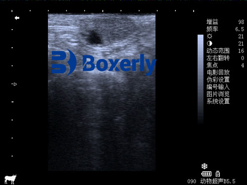
Follicular cysts, thin‑walled and anechoic on ultrasonography, with minimal luteal tissue.
-
Luteal cysts, thicker‑walled structures containing luteal tissue and often higher progesterone levels.
Cows with ovarian cysts may display irregular estrus, silent heats, or prolonged anestrus, leading to extended calving intervals and economic losses due to delayed pregnancies and increased culling.
Pathophysiology and Risk Factors
The etiology of cystic ovarian disease is multifactorial:
-
Metabolic stress in early lactation, including negative energy balance, impairs hypothalamic–pituitary–ovarian axis function.
-
High milk yield, genetic predisposition, and breed influence incidence.
-
Postpartum inflammation or uterine disorders can alter gonadotropin release, preventing normal follicular rupture.
-
Seasonality and nutrition, such as phytoestrogen intake, modulate cyst formation risk.
Understanding these risk factors facilitates herd‑level management to reduce cyst prevalence.
Ultrasonography: A Non‑Invasive Diagnostic Tool
Transrectal ultrasonography using a 7.5 MHz linear probe has become the gold standard for ovarian evaluation. This non‑invasive method offers:
-
Real‑time imaging of ovarian structures and uterine tone.
-
Measurement of cyst diameter, wall thickness, and internal echogenicity.
-
Differentiation between follicular and luteal cysts by assessing wall characteristics and blood flow patterns using Doppler modes.
Regular scanning at 10‑ to 14‑day intervals in early lactation enables early detection and timely treatment.
Scanning Protocol and Interpretation
-
Preparation: Restrain cow in chute; ensure rectum is emptied and probe lubricated.
-
Probe placement: Introduce probe transrectally, identify uterine horns, then ovaries.
-
Image acquisition: Record size (≥ 25 mm qualifies as cyst), wall thickness (≤ 3 mm for follicular, > 3 mm for luteal), and presence of corpus luteum or multiple follicles.
-
Doppler assessment: Evaluate blood flow to differentiate luteal cysts (increased vascularity) from follicular (low flow).
Correct interpretation guides therapeutic choices and breeding decisions.
Impact on Fertility and Herd Economy
Ovarian cysts prolong the calving‑to‑conception interval by 20–50 days on average, reducing lifetime productivity of affected cows. Treatment and monitoring incur veterinary costs, while reduced milk yield during anestrus and higher culling rates amplify economic losses. Timely ultrasonographic diagnosis and intervention can shorten cyst duration and restore cyclicity, recovering both reproductive performance and profitability.
Treatment Strategies
Treatment aims to induce luteinization of follicular cysts and restore normal cycles:
-
GnRH analogs (e.g., buserelin) to trigger luteinizing hormone surges and luteinization.
-
PGF₂α administration 7 days post‑GnRH to regress luteal tissue in luteal cysts.
-
Ovsynch protocols, combining GnRH/PGF₂α/GnRH, offer fixed‑time AI options without need for heat detection.
-
Supportive measures: correcting energy balance, alleviating stress, and improving body condition.
Ultrasonography is used pre‑ and post‑treatment to confirm cyst resolution and ovulation.
Case Study: High‑Yield Herd Intervention
A 120‑cow Holstein herd experiencing extended days open underwent screening at 30 and 45 days in milk. Ultrasonography identified ovarian cysts in 18 cows (15 %). A modified Ovsynch protocol was implemented:
-
Day 0: GnRH
-
Day 7: PGF₂α
-
Day 9: GnRH + fixed‑time AI
Follow‑up scans at Day 17 confirmed ovarian luteal structures in 16 cows, and conception rate improved by 45 % over untreated cohorts. Economic modeling projected a return on investment within one lactation cycle.
Prevention and Herd Management
Preventing ovarian cysts relies on minimizing risk factors:
-
Optimize transition cow diet to prevent negative energy balance.
-
Monitor body condition; avoid extremes.
-
Implement regular reproductive health screening, with ultrasonography as key component.
-
Genetic selection against cystic predisposition, removing sires whose daughters have high cyst incidence.
Comprehensive herd health protocols integrating ultrasonography can markedly reduce cystic ovarian disease prevalence.
Training and Technology Adoption
Successful implementation of ultrasonographic programs requires:
-
Training farm personnel in probe handling, image interpretation, and record‑keeping.
-
Investment in portable ultrasonography units with Doppler capabilities, suitable for on‑farm use.
-
Standardized protocols to ensure consistency across operators and herds.
Collaborations with veterinary specialists and continuing education foster high proficiency and maximize technology benefits.
Conclusion
Cystic ovarian disease poses a significant challenge to reproductive performance in high‑producing dairy cows. Veterinary ultrasonography—offering timely, précis, and non‑invasive diagnosis—has become indispensable for identifying ovarian cysts, guiding targeted therapies, and informing herd management decisions. By integrating ultrasonographic screening into routine reproductive programs, dairy producers can detect cysts early, implement effective treatments, and optimize fertility outcomes. Ultimately, this technology not only enhances individual cow productivity but also contributes to the economic sustainability of modern dairy operations.
References
-
Dinkissa AF. Review on Ovarian Cysts in Dairy Cattle, Its Treatment and Prevention. Int J Educ Appl Sci Res. 2022;9(1):1–16. DOI:10.5281/zenodo.7090356. Available at: http://doi.org/10.5281/zenodo.7090356
-
Opsomer G, et al. Reproductive performance in dairy cows with cystic ovarian disease. Theriogenology. 2018;112:11–18. Available at: https://www.ncbi.nlm.nih.gov/pmc/articles/PMC6068299/
-
Johnson AD, Coates G. Ovarian cysts: an anovulatory condition in dairy cattle. J Dairy Sci. 2005;88(6):1973–1978. Available at: https://pmc.ncbi.nlm.nih.gov/articles/PMC7653308/
-
Pacheco LSF, et al. Color Doppler ultrasonography in differentiating follicular and luteal cysts in dairy cattle. J Dairy Res. 2023;90(2):125–132. Available at: https://www.sciencedirect.com/science/article/pii/S0022030223001054
-
Yimer NA, Ghafoorunissa K. Ultrasound and progesterone monitoring of ovarian follicular cysts in dairy cattle. Anim Reprod Sci. 2006;92(1–2):18–31. Available at: https://www.sciencedirect.com/science/article/abs/pii/S0007193505802027
-
Menchaca A, Rubianes E. Efficiency of a modified ovulation synchronization protocol in dairy cows. Animals (Basel). 2024;15(7):995. DOI:10.3390/ani15070995. Available at: https://www.mdpi.com/2076-2615/15/7/995