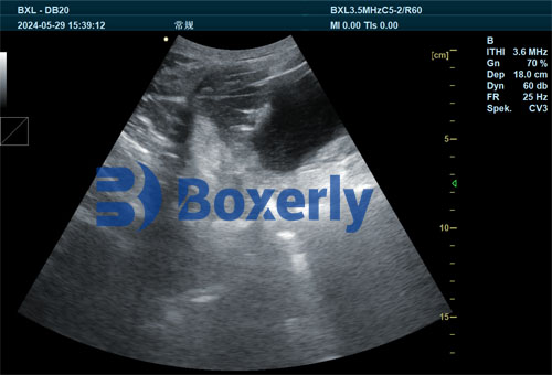
In modern veterinary diagnostics, ultrasound imaging has become a cornerstone technology for evaluating internal structures in animals, especially in livestock such as cattle, porcins, and sheep. Veterinary ultrasound is prized for being non-invasive, real-time, and highly adaptable across species. Toutefois, its clinical effectiveness relies heavily on the quality of the images produced.
Ces dernières années, with the rapid advancement of computer science and digital imaging technology, ultrasound image enhancement has emerged as a vital field. Particularly in bovine reproductive diagnostics, enhanced ultrasound images allow veterinarians to better differentiate between normal and pathological tissues, improving both diagnosis and treatment planning. Dans cet article, we will explore the theoretical principles behind veterinary ultrasound Amélioration de l’image, how it is applied in practice, and how foreign researchers and veterinarians interpret its value in clinical settings.
Why Ultrasound Image Enhancement Matters
Ultrasound imaging involves transmitting high-frequency sound waves into the animal’s body and interpreting the reflected signals to construct an image. Toutefois, échographie vétérinaire images are prone to several limitations:
-
Low contrast between soft tissues
-
Presence of speckle noise
-
Operator-dependent variability
-
Limited spatial resolution
Veterinarians and researchers around the world have identified these limitations as key barriers to accurate diagnosis, especially in early pregnancy detection, ovarian evaluation, and fetal development monitoring in cows. Consequently, ultrasound image enhancement focuses on improving clarity, removing irrelevant noise, and emphasizing diagnostic features that matter most to clinicians.
The Core Principles of Ultrasound Image Enhancement
In simple terms, ultrasound image enhancement refers to the use of digital processing techniques to make important features in the image more visible while minimizing unimportant or misleading signals. According to leading imaging researchers in Europe and North America, two main objectives guide this process:
-
Visual enhancement for direct observation: The processed image should appear clearer and more detailed to the naked eye, enabling veterinarians to interpret the results confidently in real time.
-
Structural enhancement for computational analysis: Enhanced images should also support quantitative analysis, such as measuring follicular diameter, endometrial thickness, or fetal size, especially in AI-based diagnostic systems.
Key Technologies and Techniques in Image Enhancement
Several techniques are commonly used to enhance veterinary ultrasound images. These techniques, adapted from human medical imaging, have been modified to address the specific challenges of animal physiology and veterinary workflows.
-
Speckle Reduction Algorithms
Speckle is a granular noise pattern that occurs due to the constructive and destructive interference of ultrasound waves. It can obscure fine details in soft tissue and reduce image readability. Adaptive filters, such as anisotropic diffusion and wavelet thresholding, have been widely used in both American and European veterinary hospitals to reduce speckle without blurring critical anatomical edges.
-
Edge Enhancement
In veterinary practice, being able to visualize the border of a follicle, corpus luteum, or uterine horn is critical. Edge enhancement algorithms, including Sobel and Laplacian operators, increase contrast at the boundaries of structures. In bovine reproduction management, this allows better tracking of changes in ovarian morphology or uterine wall integrity.
-
Histogram Equalization
This technique redistributes pixel intensity values to improve the overall contrast of the image. Foreign researchers have found histogram equalization particularly useful for detecting subtle tissue changes, such as distinguishing between healthy and cystic ovarian follicles in vaches laitières.
-
Region-of-Interest (ROI) Emphasis
Veterinarians are often most interested in specific areas, such as the fetus during pregnancy checks or the endometrium during estrus cycle monitoring. By isolating and enhancing the ROI using segmentation algorithms, practitioners can focus on diagnostic details while filtering out irrelevant background structures.
Global Applications and Perspectives
Veterinarians across the globe have recognized the benefits of image enhancement, especially in regions with large-scale livestock operations. Par exemple:
-
In Canada and the United States, large dairy farms utilize enhanced ultrasound to monitor reproductive efficiency, reducing open days and improving calving intervals. The emphasis is on speed and precision, and enhanced images help AI technicians make faster, more accurate decisions.
-
In European countries like Germany and the Netherlands, where animal welfare standards are stringent, enhanced ultrasound is used not only for diagnostics but also for monitoring post-treatment recovery. Veterinarians value the improved detail in uterine and ovarian structures that image enhancement provides.
-
In developing countries, mobile ultrasound systems equipped with real-time image enhancement are enabling veterinarians to offer higher-quality diagnostics in remote rural settings. These tools improve access to reproductive healthcare for livestock herders who otherwise might rely on visual or manual palpation alone.
Case Study: Bovine Reproductive Imaging
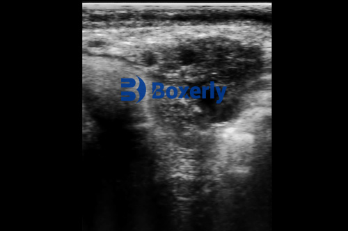
Let’s take the example of a reproductive ultrasound scan in a Holstein cow. The raw image might show a low-contrast view of the uterus and ovaries, with various echogenic (bright) and anechoic (dark) areas. Without enhancement, it may be difficult to distinguish a corpus luteum from surrounding tissue or to detect early pregnancy.
By applying enhancement techniques such as speckle noise reduction and edge detection, the image becomes clearer. The corpus luteum now appears as a sharply bordered, hypoechoic region, while the uterine horns show greater texture detail. This allows the veterinarian to confidently determine reproductive status and recommend follow-up interventions.
Subjective Evaluation and Practical Considerations
Unlike laboratory tests, the effectiveness of ultrasound image enhancement often depends on the subjective interpretation of the veterinarian. As many foreign experts have pointed out, there is no universal standard for what constitutes a “perfect” enhanced image. The value of enhancement lies in whether it helps the human eye distinguish between normal and abnormal structures.
Par exemple, a well-enhanced image for evaluating pregnancy may not be suitable for examining an inflamed ovary. En outre, excessive enhancement can introduce artifacts, leading to misinterpretation. This is why image enhancement must be used judiciously and always in conjunction with the clinical context.
Veterinarians from Australia and New Zealand emphasize the importance of training in ultrasound image interpretation. They advocate for integrating image enhancement education into veterinary school curricula to ensure that future practitioners can make the most of this technology.
Role in AI and Future Directions
As artificial intelligence (IA) gains traction in veterinary medicine, high-quality ultrasound images are becoming essential training data for machine learning models. Enhanced images, with clear boundaries and labeled regions, provide the perfect input for AI algorithms tasked with identifying pathological changes automatically.
Several research groups in the U.S. and China are developing AI-powered systems that can detect ovarian cysts or predict the likelihood of successful insemination based on enhanced ultrasound images. The synergy between enhancement and AI holds great promise for transforming livestock reproduction management.
Conclusion
Veterinary ultrasound image enhancement is not just a technical improvement—it is a clinical necessity. By improving clarity, suppressing noise, and highlighting diagnostically relevant regions, image enhancement empowers veterinarians to make faster, more accurate, and more confident decisions. Although enhancement techniques vary based on region, purpose, and species, the universal goal is the same: to make the invisible visible and support the health and productivity of animals.
As veterinary medicine continues to adopt AI and data-driven diagnostics, the role of image enhancement will only grow. Veterinarians, farmers, and technologists must work together to ensure that the full potential of this powerful tool is realized in both high-tech labs and rural barns alike.